Elevated serum gamma globulins in apparently healthy Nigerians living in Ogbomoso: a possible manifestation of phagocytic dysfunction
Abstract
Background: Serum protein abnormalities, particularly elevated gamma globulins (hypergammaglobulinemia, HGG), have been reported in apparently healthy Nigerians living in Ogbomoso and elsewhere. Since the mechanisms for this phenomenon have not been fully substantiated, we hypothesized that impaired neutrophil phagocytosis could contribute to this condition.
Methods: Healthy humans exhibiting HGG were identified using serum protein electrophoresis performed on cellulose acetate gel in barbital buffer (pH 8.6). GelQuant image analysis and quantitation software were further employed to quantify the gamma globulin fraction. Neutrophils were isolated from K3EDTA anticoagulated peripheral blood using Histopaque neutrophil isolation reagent. Neutrophil phagocytic activity was analyzed using a non-subjective commercial colorimetric phagocytosis assay kit.
Results: The purity and viability of isolated neutrophils were approximately 94% and 92%, respectively. Ex-vivo phagocytic activity of neutrophils isolated from apparently healthy subjects exhibiting HGG, expressed as a percentage of the average absorbance of the control group, was 48.1 ± 8.6% which was significantly lower (p < 0.05) compared to the controls (98.9 ± 14.3%).
Conclusion: Since neutrophils play crucial roles in innate immune responses, impairment of neutrophil phagocytic activity may lead to persistent antigenic stimulations of the adaptive immune system. This could in turn orchestrate gamma globulins expression leading to HGG.
Statement of novelty: We demonstrated reduced neutrophil phagocytic activity as a possible basis for hypergammaglobulinemia in healthy Nigerians, perhaps for the first time.
Introduction
Phagocytosis is defined as a receptor-mediated process in which targeted particles are engulfed and degraded. Neutrophils are the predominant leukocytes in peripheral blood and from there, they are mobilized to the sites of infection (Teng et al. 2017; Leach et al. 2019). Although neutrophils do not possess the properties for adaptive recognition of antigens, nevertheless, they mediate early innate immune responses to infection and display the capacity to modulate the adaptive immune response (Ley et al. 2018; Papayannopoulos 2018; Silvestre-Roig et al. 2019; Rosales 2020). Neutrophils are also important sources of pro- and anti-inflammatory cytokines, thus participating in host defenses through a variety of phenotypically simple but mechanistically complex processes (Tamassia et al. 2018; Gideon et al. 2019; Kumar 2020). Other neutrophil functions include chemotaxis, respiratory burst activity, direct bacterial killing, and antibody-dependent cell-mediated cytotoxicity, and most of these functions can be demonstrated in vitro (Lehman and Segal 2020).
Immunoglobulins are glycoproteins produced by plasma cells and are key effector factors of the adaptive immune system. The Fc regions of IgG molecules are involved in complement and antibody-dependent phagocytosis (Quast et al. 2017). IgG is the most abundant immunoglobulin and therefore constitutes most of the gamma region in serum protein electrophoresis (Adedeji et al. 2014). Hypergammaglobulinemia (HGG) is found in many situations. In the setting of primary immunodeficiency, HGG could, for instance, be related to a defect in T cell function, liver diseases, malignancies, autoimmune diseases, and infections (Upton 2014). Although immunodeficiency may be associated with a significant decrease in plasma IgG, IgM, or IgA isotypes, some immunodeficiency conditions with normal or elevated levels of immunoglobulins have been documented (Conley et al. 1999; Adedeji et al. 2014; Pimenta et al. 2019). Furthermore, neutrophil functions are reduced in the immunodeficiency state, such as in HIV-infection (Dantas et al. 2015). Some immunodeficiency diseases are very well described as having high levels of a particular immunoglobulin. For instance, hyper IgM usually results from the impaired ability of B cells to undergo immunoglobulin class-switching, while IgE usually predominates in hyper IgE syndrome as well as other conditions (Lo et al. 2013).
Previous studies have shown that some healthy humans have exhibited HGG (Buadi et al. 2011; Adedeji et al. 2015). This phenomenon was first recognized in Nigerians living in Britain as far back as the 1950s (Schofield 1957), but the precise immunological basis for this condition is still emerging. Since neutrophils are crucial in the first line of defense and immunoglobulins are expressed following antigenic stimulation, we hypothesized that impaired neutrophil phagocytic function would contribute to this phenomenon. Although phagocyte dysfunction has been reported in pathologic conditions (Carneiro et al. 2012; Teng et al. 2017), data on phagocytic activity of neutrophils isolated from peripheral blood of apparently healthy individuals exhibiting HGG are scarce. Thus, we investigated whether neutrophil dysfunction is associated with HGG in apparently healthy individuals, as this could provide a possible basis for this phenomenon.
Methods
Subject selection
This is a cross-sectional study where 100 healthy undergraduates in the Faculty of Basic Medical Sciences, Ladoke Akintola University of Technology, Ogbomoso (Supplementary Material1), Nigeria, who met the selection criteria were recruited. Volunteers were required to complete a structured questionnaire. Those with a medical history that could influence the results at the time of enrolment were excluded from the study. Blood samples were collected from the participants after overnight fasting. The serum was separated by centrifugation and stored at −20 °C. The study was approved by the Ethical Committee of the Faculty of Basic Medical Sciences and informed consent was obtained from all volunteers before the study was initiated.
Serum protein electrophoresis (SPE)
SPE was performed on cellulose acetate gel in barbital buffer (pH 8.6) at 20 V/cm for 25 minutes. The Helena electrophoresis system (Helena Laboratories, Beaumont, TX) was used to identify participants exhibiting HGG. SPE was performed according to the recommendations provided by the manufacturer. The separated fractions were fixed in 5% acetic acid and visualized with Ponceau S stain. The electropherogram was independently examined by 2 of the authors (ALA and IES) to identify participants exhibiting HGG and any discrepancies were resolved by consensus. To quantitatively define HGG, serum protein fractions were measured both in HGG participants and the controls using GelQuant image analysis and quantitation software, as described by Khakabimamaghani et al. (2013). Absolute protein fractions were calculated from total protein concentration, determined using the Biuret method of Weichselbaum (1946). Serum albumin concentration was determined by the method of Doumas et al. (1971).
Serum immunoglobulin estimation
Serum immunoglobulin (total IgG, IgM, and IgA) concentrations were determined by Mancini single radial immunodiffusion (Mancini et al. 1965), modified by Liofilchem S.R.L., Italy.
Liver and renal function assessment
Renal and liver function of the HGG subjects were assessed to rule out liver diseases. Serum aspartate aminotransferase (AST) and alanine aminotransferase (ALT) activities were determined by the colorimetric method as described by Reitman and Frankel (1957). Serum alkaline phosphatase (ALP) activities were measured by the colorimetric method as described by Plummer (1978) with phenolphthalein monophosphate used as substrate. Serum urea and creatinine concentrations were determined by modified Berthelot reaction (Burtis and Ashwood 1999) and modified Jaffe reaction (Mazzachi et al. 2000) methods, respectively. The reagents used for the enzyme activity, urea, and creatinine assays were obtained from assay kits of Randox Laboratories Ltd., UK.
Isolation of neutrophils
Neutrophils were isolated from whole blood using neutrophil isolation reagent, obtained from Cayman Chemical, USA. The reagent was stored and used according to modified manufacturer instructions. To prevent neutrophil activation during the separation procedure, EDTA anticoagulated blood was used and the washing PBS did not contain Ca2+/Mg2+, since these ions have been shown to prime cells (Oh et al. 2008; Freitas et al. 2008). Briefly, 5 ml of K3EDTA anticoagulated blood diluted 1:2 in cell-based assay buffer (Item No 10009322) was layered on 3.33 mL of cell-based assay neutrophil isolation Histopaque reagent (Item No 600612), in a 15 mL conical tube. It was centrifuged at 500 × g for 25 minutes at 26 °C. The top layers were carefully removed and 10 ml of Red Blood Cell Lysis Buffer (Item No 601077) was added for 10 minutes to lyse the red blood cells. The neutrophil fraction was pelleted at 1200 rpm for 10 minutes. The reddish supernatant was carefully aspirated and the remaining pellet was washed twice in 1.66 ml of RPMI containing 1% BSA. Resuspension of pellets was performed slowly and rested as recommended by Kutscher et al. (2013). The isolated neutrophils were suspended in 5 mL RPMI containing 1% BSA and rested for 10 minutes at 26 °C.
Assessment of viability and purity of isolated neutrophils and phagocytic activity assay
The purity and viability of the isolated neutrophils were evaluated by Leishman’s staining and trypan blue dye exclusion method, respectively, as described by Joshi et al. (2020). The neutrophil phagocytic capacity was performed using zymosan (Saccharomyces cerevisiae) commercially prepared from yeast cell wall, which consists of protein-carbohydrate complexes (purchased from Cell Biolabs, USA). The CytoSelectTM 96-well phagocytosis assay that uses prelabelled zymosan particles as a phagocytosis pathogen was employed in the study. The phagocytosed zymosan particles were determined by measuring the absorbance of each well at 405 nm. The results were expressed as a percentage of the average absorbance of the control group.
Statistical analysis
Descriptive analysis and student t-test were used for the comparison of data. Spearman correlation was used to test the association between variables, using Graphpad® 5 software (San Diego, CA). P-values <0.05 were considered significant.
Results
Study participants
Seven (7%) of a total of 100 healthy participants initially recruited were identified to exhibit HGG. Seven participants exhibiting normal gamma globulin bands were randomly selected as controls. The renal and liver indices and other details are presented in Table 1.
Table 1:
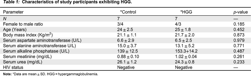
Note: *Data are mean ± SD. HGG = hypergammaglobulinemia.
Serum protein pattern and neutrophil phagocytic activity
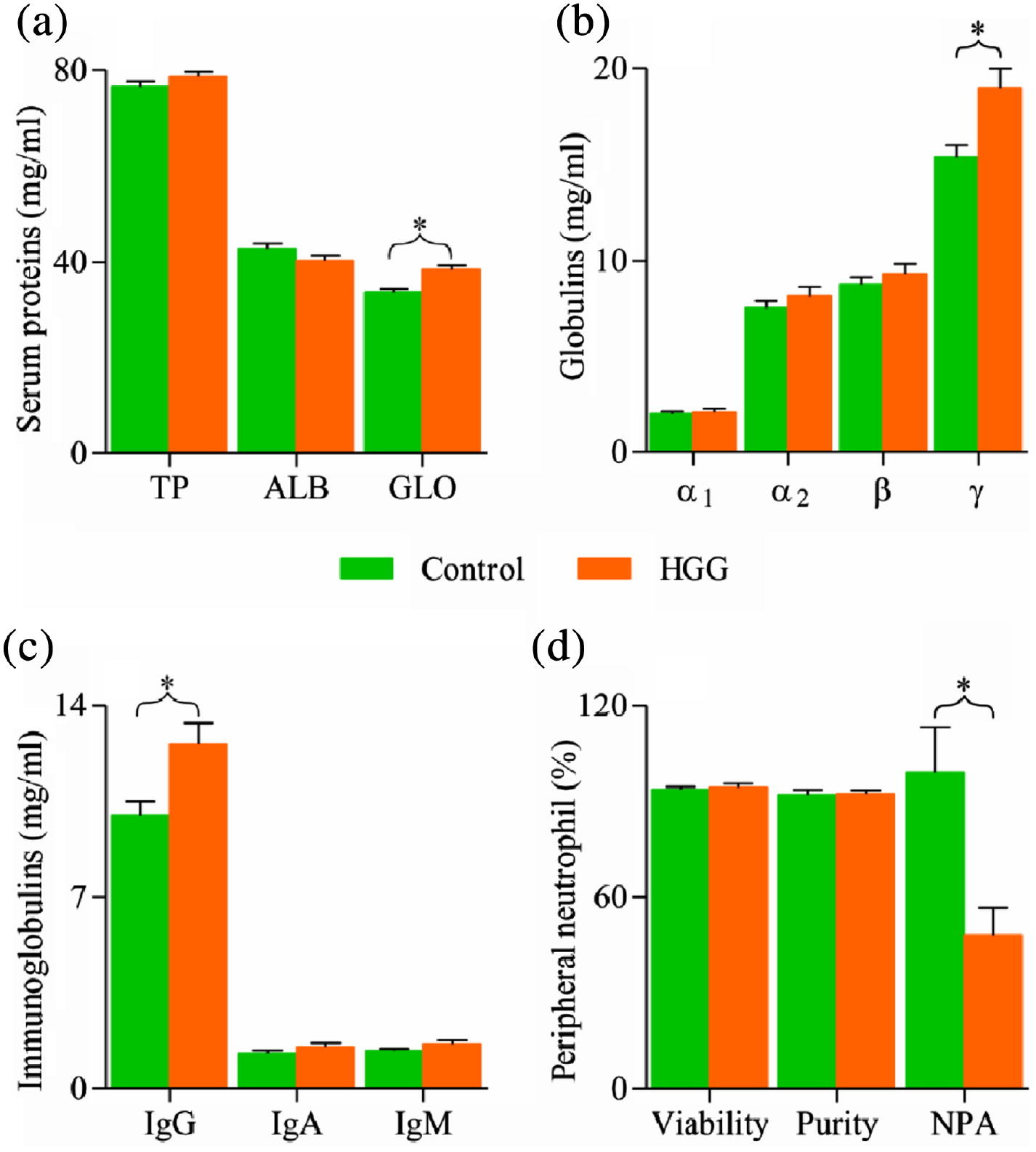
Serum proteins and albumin concentrations in HGG were not significantly (p < 0.05) different from the controls whereas the concentrations of IgG and gamma globulin were significantly (p < 0.05) higher in HGG compared with the control. The changes in other immunoglobulin concentrations were not significant (p > 0.05). The purity and viability (94% and 92%, respectively) of isolated neutrophils were adequate for the in vitro phagocytosis assay. Figure 1 shows that the phagocytic activity of neutrophils isolated from apparently healthy participants exhibiting HGG is significantly lower than the controls (p < 0.05).
Figure 1:
Association between neutrophil phagocytic activity and serum immunoglobulins
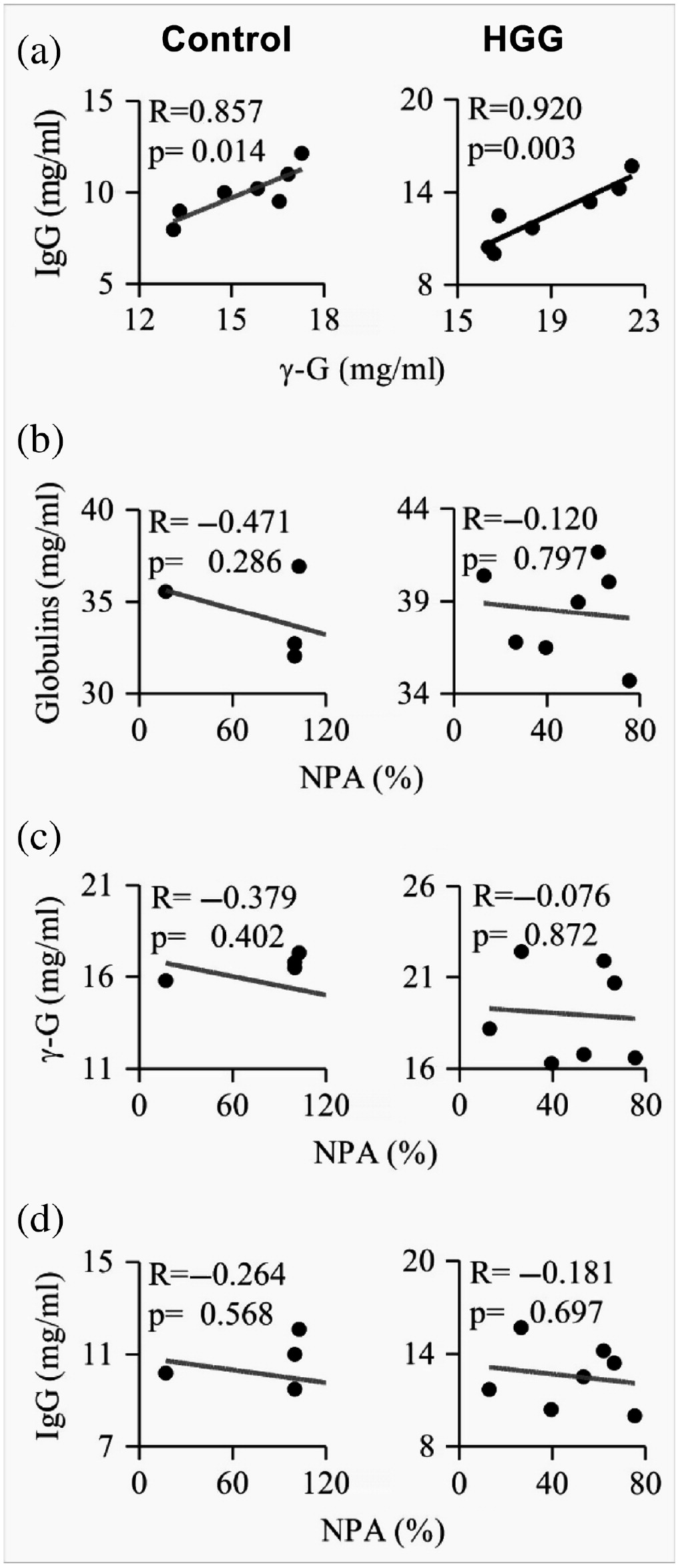
Figure 2 shows the correlation of neutrophil phagocytic activity (NPA) with immunoglobulins in HGG and the controls. IgG positively correlated with gamma globulin (R = 0.920; p = 0.003) and (R = 0.867; p = 0.014), respectively, in HGG and controls. No significant correlation was observed between neutrophil phagocytic activity and gamma globulin, IgG, in both the HGG and control group (p > 0.05).
Figure 2:
Discussion
In this report, we demonstrated reduced phagocytotic activity in neutrophils isolated from apparently healthy participants exhibiting HGG compared with the controls. We first identified healthy participants showing HGG by SPE, and subsequently confirmed HGG by quantifying the gamma globulin band using GelQuant image analysis and quantitation software (Khakabimamaghani et al. 2013).
Since HGG has been reported in patients with chronic liver disease (Fallatah and Akbar 2010), we ruled out this condition by studying the liver and renal indices such as plasma AST, ALT, ALP, urea and creatinine. The liver and renal indices and HIV status reports confirmed that HGG is not due to underlying liver disease in the study participants. Other demographic parameters in both HGG and control participants were essentially the same (p > 0.05) (Table 1).
SPE is invaluable in health and diseases. It has 3 traditional applications; the first application is in the identification and characterization of serum protein abnormalities, the second application is in identifying the transition from monoclonal gammopathy of undetermined significance to multiple myeloma, and the third application is in disease activity or treatment in multiple myeloma (Katzmann et al. 1997). The procedure is now very useful in identifying healthy humans exhibiting HGG. Different varieties of SPE have been reported depending on the nature of the support medium employed. The 3 commonly used support media, cellulose acetate, agarose, and capillary, each have different specificity and sensitivity in determining serum protein abnormalities (Katzmann et al. 1997). It is noteworthy that the cellulose acetate support medium used in this study has been reported to have excellent specificity.
The concentration of serum gamma globulin and IgG were significantly (p < 0.05) higher in the HGG group compared to the control. Immunofixation electrophoresis (IFE) is a research tool for identifying the electrophoretic mobility of serum protein fractions in blood and other biological fluids. With the use of IFE, several proteins have been identified to migrate in the gamma band of SPE. Notably amongst them is IgG. Others are IgA, IgM, and complement C3 (Yousif et al. 2018). In this study, gamma globulin and total IgG were positively correlated and it is evident from previous reports that the major component of the gamma band is IgG. Phagocytic activity of neutrophils was demonstrated to be significantly reduced in participants exhibiting HGG. Anomalies in neutrophil phagocytic function have been observed in several common medical and surgical conditions (Engelich et al. 2001). Its occurrence among participants exhibiting HGG is still emerging.
As is generally known, phagocytosis is initiated by the interaction of phagocytic receptors with ligands on the surface of target microbes. Then, receptors aggregate to initiate signaling pathways that regulate the actin cytoskeleton, so that the phagocyte can produce membrane protrusions to engulf microbes. Lastly, the microbes are encircled in a new vesicle that protrudes out from the plasma membrane. The impairment in NPA may result from 1 or more of the many well-defined and complex processes that have been systematically divided into 4 principal steps: recognition of pathogens, activation of the internalization process, formation of the phagosome, and phagolysosome maturation. These processes have been recently reviewed (Nordenfelt and Tapper 2011; Rosales and Uribe-Querol 2017; Liew and Kubes 2019). Further studies will be needed to demonstrate precisely the molecules that are defective in this condition. However, phagocytic defects are generally a consequence of impaired actin polymerization around phagosomes (May and Machesky 2001; Baranov et al. 2016).
The correlation studies show that IgG is implicated in HGG and significant correlation was observed between total IgG and gamma globulins (Figure 1), and these may be expressed against microbes that breached the first line of defense. Determination of specific antibodies would add an interesting dimension to this study. Furthermore, evaluation of the IgG subtype may reveal a predominant subclass in this condition. For instance, IgG1 and IgG3 subclass predominate in HIV infection and liver cirrhosis, respectively (Riggione et al. 1983; Kekow et al. 1988).
Since neutrophils play crucial roles as effector molecules in the first line of defense in humans, impairment of neutrophil phagocytic activity may favour persistent antigenic stimulation of the adaptive immune system. This in turn could orchestrate gamma globulin expression leading to HGG. Consequently, there might be a reduced ability of neutrophils, in this subset of young individuals, to combat microbial infections. However, since no peculiar symptom is associated with reduced neutrophil phagocytosis in apparently healthy individuals considered in this study, this phenomenon could be termed reduced neutrophil phagocytic activity of undetermined significance.
Conflict of interest
The authors declare no conflict of interest. This research did not receive any specific grant from funding agencies in the public, commercial, or not-for-profit sectors.
Author’s contribution
ALA conceived and designed the study, and collected data. JAB contributed to laboratory reagents and analysis tools. JD and IOB collected data and performed data analysis. IES drafted and revised the manuscript. OGA supervised laboratory analysis and revised the manuscript.
Funding
The research was funded by the authors.
Footnote
1
Supplementary data are available with the article through the journal Web site at http://lymphosign.com/doi/pdf/10.14785/lpsn-2021-0024.
REFERENCES
Adedeji A.L., Adenikinju R.O., Ajele J.O., and Olawoye T.L. 2014. Serum protein electrophoresis under effective control of HIV-1 disease progression. EXCLI J. 13: 761–771.
Adedeji A.L., Faniran O.G., and Olawoye T.L. 2015. Immunologic characteristics of apparently healthy Nigerians exhibiting abnormal SPE: A preliminary study. RRJoI. 5(3): 1–6.
Baranov M.V., Revelo N.H., Dingjan I., Maraspini R., Ter Beest M., Honigmann A., and van den Bogaart G. 2016. SWAP70 organizes the actin cytoskeleton and is essential for phagocytosis. Cell Rep. 17(6): 1518–1531.
Buadi F., Hsing A.W., Katzmann J.A., Pfeiffer R.M., Waxman A., Yeboah E.D., Biritwum R.B., Tettey Y., Adjei A., Chu L.W., DeMarzo A., Netto G.J., Dispenzieri A., Kyle R.A., Rajkumar S.V., and Landgren O. 2011. High prevalence of polyclonal hypergamma-globulinemia in adult males in Ghana, Africa. Am. J. Hematol. 86(7): 554–558.
Burtis, C.A., and Ashwood, E.R. 1999. Tietz textbook of clinical chemistry. Philadelphia.
Carneiro V.M.A., Bezerra A.C.B., Guimarães M.D.C.M., and Muniz-Junqueira M.I. 2012. Decreased phagocytic function in neutrophils and monocytes from peripheral blood in periodontal disease. J. Appl. Oral. Sci. 20(5): 503–509.
Conley M.E., Notarangelo L.D., and Etzioni A. 1999. Diagnostic criteria for primary immunodeficiencies. Clin. Immunol. 93(3): 190–197.
Dantas E.O., Aranda C.S., Rêgo Silva A.M., Tavares F.S., Severo Ferreira J.F., de Quadros Coelho M.A., de Siqueira Kovalhuk L.C., Roxo Júnior P., Toledo E.C., Porto Neto A.C., de Sousa Vieira H.M., Takano O.A., Nobre F.A., Sano F., Nudelman V., de Farias Sales V.S., Silva Segundo G.R., Villar Guedes H.T., Félix E., Marques S.M., and Costa Carvalho B.T. 2015. Doctors’ awareness concerning primary immunodeficiencies in Brazil. AllergolImmunopathol. 43(3): 272–278.
Doumas B.T., Watson W.A., and Biggs H.G. 1971. Albumin standards and the measurement of serum albumin with bromocreasol green. Clin. Chim. Acta. 31(1): 87–96.
Engelich G., Wright D.G., and Hartshorn K.L. 2001. Acquired disorders of phagocyte function complicating medical and surgical illnesses. Clin. Infect. Dis. 33(12): 2040–2048.
Fallatah H.I. and Akbar H.O. 2010. Elevated serum immunoglobulin G levels in patients with chronic liver disease in comparison to patients with autoimmune hepatitis. Libyan. J. Med. 5.
Freitas M., Porto G., Lima J.L., and Fernandes E. 2008. Isolation and activation of human neutrophils in vitro. The importance of the anticoagulant used during blood collection. Clin. Biochem. 41(7–8): 570–575.
Gideon H.P., Phuah J., Junecko B.A., and Mattila J.T. 2019. Neutrophils express pro- and anti-inflammatory cytokines in granulomas from Mycobacterium tuberculosis-infected cynomolgus macaques. Mucosal. Immunol. 12(6): 1370–1381.
Joshi M.B., Ahamed R., Hegde M., Nair A.S., Ramachandra L., and Satyamoorthy K. 2020. Glucose induces metabolic reprogramming in neutrophils during type 2 diabetes to form constitutive extracellular traps and decreased responsiveness to lipopolysaccharides. BiochimBiophys. Acta Mol. Basis Dis. 1866(12): 165940.
Katzmann J.A., Clark R., Wiegert E., Sanders E., Oda R.P., Kyle R.A., Namyst-Goldberg C., and Landers J.P. 1997. Identification of monoclonal proteins in serum: a quantitative comparison of acetate, agarose gel, and capillary electrophoresis. Electrophoresis, 18(10): 1775–1780.
Kekow J., Hobusch G., and Gross W.L. 1988. Predominance of the IgG1 subclass in the hypergammaglobulinemia observed in pre-AIDS and AIDS. Cancer Detect Prev. 12(1–6): 211–216.
Khakabimamaghani S., Najafi A., Ranjbar R., and Raam M. 2013. GelClust: a software tool for gel electrophoresis images analysis and dendrogram generation. Comput Methods Programs Biomed. 111(2): 512–518.
Kumar V. 2020. Phagocytosis: Phenotypically simple yet a mechanistically complex process. Int. Rev. Immunol. 39(3): 118–150.
Kutscher S., Dembek C.J., Deckert S., Russo C., Körber N., Bogner J.R., Geisler F., Umgelter A., Neuenhahn M., Albrecht J., Cosma A., Protzer U., and Bauer T. 2013. Overnight resting of PBMC changes functional signatures of antigen specific T- cell responses: impact for immune monitoring within clinical trials. PLoS ONE, 8(10): e76215.
Leach J., Morton J.P., and Sansom O.J. 2019. Neutrophils: Homing in on the myeloid mechanisms of metastasis. Mol. Immunol. 110: 69–76.
Lehman H.K. and Segal B.H. 2020. The role of neutrophils in host defense and disease. J. Allergy Clin. Immunol. 145(6): 1535–1544.
Ley K., Hoffman H.M., Kubes P., Cassatella M.A., Zychlinsky A., Hedrick C.C., and Catz S.D. 2018. Neutrophils: New insights and open questions. Sci. Immunol. 3(30): eaat4579.
Liew P.X. and Kubes P. 2019. The Neutrophil’s Role During Health and Disease. Physiol. Rev. 99(2): 1223–1248.
Lo M.S., Zurakowski D., Son M.B., and Sundel R.P. 2013. Hypergammaglobulinemia in the pediatric population as a marker for underlying autoimmune disease: A retrospective cohort study. Pediatr. Rheumatol. Online J. 11(1): 42.
Mancini G., Carbonara A.O., and Heremans J.F. 1965. Immunochemical quantitation of antigens by single radial immunodiffusion. Immunochemistry, 2(3): 235–254.
May R.C. and Machesky L.M. 2001. Phagocytosis and the actin cytoskeleton. J. Cell Sci. 114(Pt 6): 1061–1077.
Mazzachi B.C., Peake M.J., and Ehrhardt V. 2000. Reference range and method comparison studies for enzymatic and Jaffé creatinine assays in plasma and serum and early morning urine. Clin. Lab. 46(1–2): 53–55.
Nordenfelt P. and Tapper H. 2011. Phagosome dynamics during phagocytosis by neutrophils. J. Leukoc. Biol. 90(2): 271–284.
Oh H., Siano B., and Diamond S. 2008. Neutrophil isolation protocol. J. Vis. Exp. 23(17): 745.
Papayannopoulos V. 2018. Neutrophil extracellular traps in immunity and disease. Nat. Rev. Immunol. 18(2): 134–147.
Pimenta F., Palma S., Constantino-Silva R.N., and Grumach A.S. 2019. Hypogammaglobulinemia: a diagnosis that must not be overlooked. Braz. J. Med. Biol. Res. 52(10): e8926.
Plummer, D.T. 1978. An introduction to practical biochemistry, 3rd ed. McGraw - Hill book company, Maidenhead.
Quast I., Peschke B., and Lünemann J.D. 2017. Regulation of antibody effector functions through IgG Fc N-glycosylation. Cell Mol. Life Sci. 74(5): 837–847.
Reitman S. and Frankel S. 1957. A colorimetric method for the determination of serum glutamic oxalacetic and glutamic pyruvic transaminases. Am. J. Clin. Pathol. 28(1): 56–63.
Riggione O., Stokes R.P., and Thompson R.A. 1983. Predominance of IgG3 subclass in primary cirrhosis. Br. Med. J. 286(6370): 1015–1016.
Rosales C. 2020. Neutrophils at the crossroads of innate and adaptive immunity. J. Leukoc. Biol. 108(1): 377–396.
Rosales C. and Uribe-Querol E. 2017. Phagocytosis: A Fundamental Process in Immunity. Biomed. Res. Int. 2017: 9042851.
Schofield F.D. 1957. The serum protein pattern of West Africans in Britain. Trans. R. Soc. Trop. Med. Hyg. 51(4): 332–337.
Silvestre-Roig C., Fridlender Z.G., Glogauer M., and Scapini P. 2019. Neutrophil diversity in health and disease. Trends Immunol. 40(7): 565–583.
Tamassia N., Bianchetto-Aguilera F., Arruda-Silva F., Gardiman E., Gasperini S., Calzetti F., and Cassatella M.A. 2018. Cytokine production by human neutrophils: Revisiting the “dark side of the moon”. Eur. J. Clin. Invest. 48(Suppl 2): e12952.
Teng T.S., Ji A.L., Ji X.Y., and Li Y.Z. 2017. Neutrophils and immunity: From bactericidal action to being conquered. J. Immunol. Res. 2017: 9671604.
Upton J. 2014. Immunodeficiencies with hypergammaglobulinemia: A review. LymphoSign Journal, 2(2): 57–73.
Weichselbaum T.E. 1946. An accurate and rapid method for the determination of proteins in small amounts of blood serum and plasma. Am. J. Clin. Pathol. 10: 40–49.
Yousif A.M., Ablhad N.S., and Ismail P.A. 2018. Serum immunofixation electrophoresis as a diagnostic method for monoclonal gammopathies in patients with multiple myeloma. Al-Mustansiriyah J. Sci. 29(4).
Supplementary Material
File (lymphosign-2021-0024suppla.pdf)
- Download
- 40.27 KB
Information & Authors
Information
Published In

LymphoSign Journal
Volume 8 • Number 3 • September 2021
Pages: 86 - 93
History
Received: 14 August 2021
Accepted: 25 August 2021
Accepted manuscript online: 27 August 2021
Copyright
© 2021.
Authors
Metrics & Citations
Metrics
Other Metrics
Citations
Cite As
Adebayo LawrenceAdedeji, DaudaJimoh, Jelili AbiodunBadmus, Ibrahim OlabanjiBello, Ibrahim ElehaSuleiman, and Olubunmi GloriaAyelagbe. 2021. Elevated serum gamma globulins in apparently healthy Nigerians living in Ogbomoso: a possible manifestation of phagocytic dysfunction. LymphoSign Journal.
8(3): 86-93. https://doi.org/10.14785/lymphosign-2021-0024
Export Citations
If you have the appropriate software installed, you can download article citation data to the citation manager of your choice. Simply select your manager software from the list below and click Download.
There are no citations for this item
View Options
View options
Login options
Check if you access through your login credentials or your institution to get full access on this article.
