Open access

Transcervical thymic biopsy in the immunodeficient child

Publication: LymphoSign Journal
4 November 2019

Abstract

Objective: The objectives of this study are to present a case series of immunodeficient children who underwent a transcervical thymic biopsy and to describe the transcervical approach to the thymus gland.
Design: Case series.
Setting: Pediatric otolaryngology practice in an academic setting.
Patients: Consecutive sample of immunodeficient children (≤18 years old) who underwent thymic biopsies from 1996 to 2019 for the purpose of confirming or excluding profound T cell immunodeficiency.
Intervention: Diagnostic transcervical thymic biopsy.
Results: A total of 14 patients with atypical combined immunodeficiency underwent the procedure during the study period, with minimal post-operative complication. The thymus was found to be abnormal histologically in 9 children and normal in another 5 patients. In all cases, thymus morphology helped define the extent of the immunodeficiency, resulting in either supporting a decision to perform a bone marrow transplant (8 patients) or avoid this high risk procedure (3 patients).
Conclusion: Thymus biopsy is helpful in the characterization of childhood immunodeficiency and provides critical information that affects the medical management. The transcervical approach to the thymus is feasible in children and can be accomplished with minimal morbidity.
Statement of novelty: Biopsies of the thymus have assisted in the characterization of new entities of primary immunodeficiency.

Introduction

The thymus is the site of T cell education and differentiation, and thus plays an important role in the body’s defense against infections. Multi-potent stem cells originate in the bone marrow and migrate to the thymic epithelium during the first trimester of gestation. Under the influence of the thymic environment, pre-T cells proliferate and acquire the functional capacity to mediate cytotoxicity and to secrete a variety of lymphokines (Janossy et al. 1980). Abnormalities in the development of the cell-mediated immune system can lead to serious and potentially fatal infections.
Thymic biopsy is a useful diagnostic modality in the workup of a child suspected of having atypical primary immunodeficiency (Hong and Pellett 1978; Leonard et al. 1985; Joshi et al. 1986). In these cases, determining the histologic and molecular basis of the immunodeficiency may influence the therapeutic options offered to the patient. In addition, thymic biopsies contribute to the description of novel entities of immunodeficiency (such as Zap-70, CD35 and CD25 deficiencies) and thereby enhance the understanding of immune system pathology (Roifman 2005).
Thymic biopsies may be obtained trans-sternally, via a thoracotomy, thoracoscopically or by a transcervical route (Hong and Pellett 1978; Leonard et al. 1985; Joshi et al. 1986). In young children, the transcervical approach may be the least invasive technique. The objectives of this study, therefore, are to describe the transcervical approach to the thymus and to present the histopathological and immunophenotypical features of the thymus gland in a series of children with primary immunodeficiency.

Methods

The medical records of immunodeficient children (age ≤18 years) who had undergone transcervical thymic biopsies at the Hospital for Sick Children, Toronto, ON, Canada from 1996 to 2019 were reviewed. Patient demographics, co-morbidities, immune status, surgical details (duration of surgery, complications), and histopathology reports were recorded.
Approval for this study was granted by the Research Ethics Board of the Hospital for Sick Children, Toronto, ON, Canada.

Surgical technique

With the neck in a fully extended position, a horizontal incision is made approximately 1 cm above the sternal notch. The underlying strap muscles are retracted and the dissection is continued inferiorly towards the superior mediastinum in the pretracheal plane. The innominate artery and vein may be encountered during the dissection immediately deep to the thymus.
Useful landmarks that aid in the identification of the thymus include the thyro-thymic ligaments, which are fibrous bands that connect the superior poles of the thymus to the inferior poles of the thyroid gland. The thymus can also be visually differentiated from the surrounding fatty tissue since it is a deeper tan-colored structure (Figure 1).
Figure 1:
Figure 1: Intra-operative photo of transcervical diagnostic thymic biopsy, showing its relation to the trachea.
An incisional biopsy is obtained from one or both upper poles of the thymus. Hemostasis of the body of the thymus should be meticulous and include suture ligatures and bipolar cautery. Placement of a surgical drain to prevent hematoma formation is not routinely employed if the surgical area is dry. The retrieved thymus tissue is then placed in normal saline and dispatched immediately to the pathology and immunology laboratories.

Results

Patients and surgical procedure

Fourteen children underwent a transcervical thymic biopsy during the study period. Seven patients were female, and 7 were male. The median age of patients at surgery was 1 year and 6 months (range: 2 months–16 years and 8 months of age), with 9 of the children being <2 years old. The more common presenting signs and symptoms of these patients are summarized in Table 1. Other presentations included cutaneous lesions, sepsis, hepatosplenomegaly, diffuse adenopathy, hypothyroidism, bronchiectasis and anemia.
Table 1:
Table 1: Common presenting signs and symptoms in immunodeficient children undergoing transcervical thymic biopsies.

Note: URTI, upper respiratory tract infection; LRTI, lower respiratory tract infections; AOM, acute otitis media; UTI, urinary tract infection.

Nine patients had unilateral biopsies of the superior poles of the thymus, while 5 patients had both lobes of their thymus biopsied. In the 7 patients who did not have concurrent surgical procedures, the mean procedure time for the thymic biopsy was 68 minutes (range 48–100 minutes). A surgical drain was inserted in 9 patients. The only post-operative complication was 1 wound infection which was treated successfully with intravenous antibiotics.
The biopsy samples were subjected to histopathological and immunohistochemical analyses and are summarized in Table 2.
Table 2:
Table 2: Summary of histopathological and immunohistochemical analyses.

Note: SCID, severe combined immunodeficiency; CID, combined immunodeficiency; PCP, Pneumocystis carinii pneumonia; Dysplastic thymus, loss of cortico medullary distinction and lack of Hassall’s corpuscles.

Evaluation of the immune system

All patients were investigated for a putative profound T cell deficiency. Mitogenic responses to phytohemagglutinin and anti-CD3 antibody were consistently depressed in all patients. However, the number of circulating T cells, with the exception of 2 patients (p8, p14) were not markedly reduced as expected in typical Severe Combined Immune Deficiencies (SCID), hence the need for assessing the thymus. In spite of the reduced function, 4 patients had normal numbers of circulating T cells, while 8 others had reduced but not absent numbers (>1000 CD3+ cells).
Assessment of humoral immunity revealed that all patients but 2 (p5, p12) had abnormal immunoglobulin and (or) antibody levels. The genetic abnormality underlying the immunodeficiency was identified only in 3 patients.

Thymus pathology

The thymus gland was found to be normal in 5 patients (Table 2). In 2 of these 5 patients (p10, p12), the biopsy results helped to avoid bone marrow transplantation (BMT). In the other 3 patients, the pathology findings were useful in further defining the diagnosis and identifying novel immunodeficiencies.
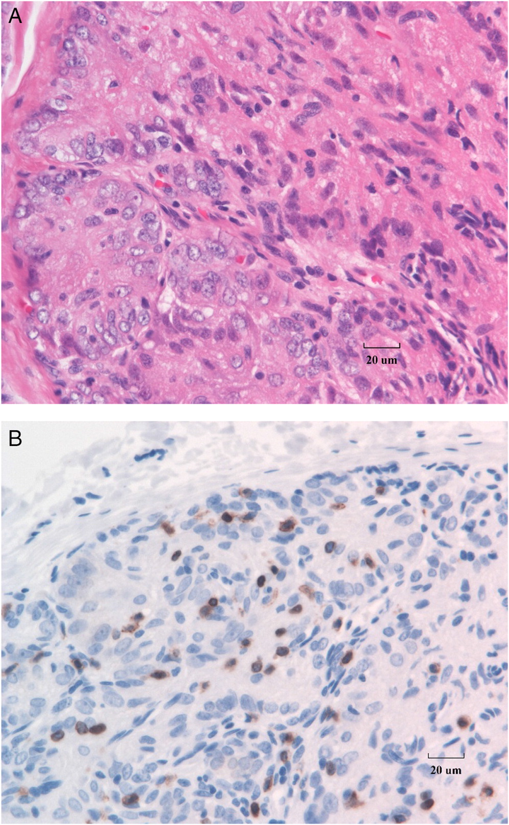
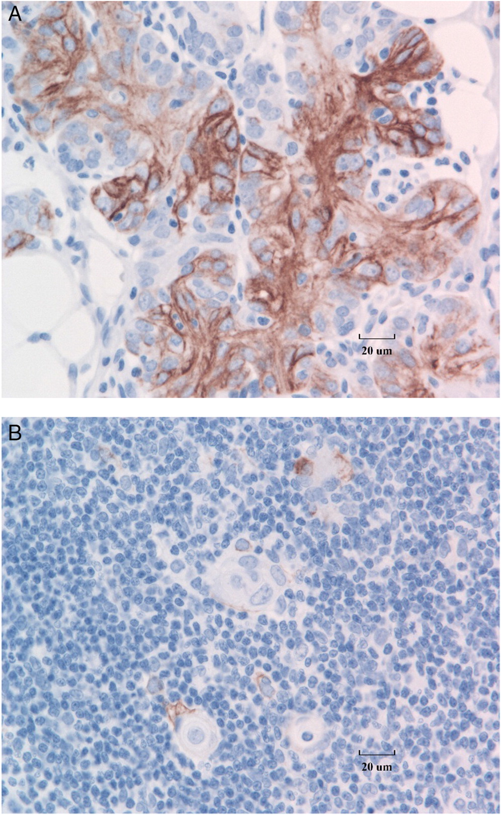
The rest of the patients had morphological abnormalities of the thymus gland. Classic dysplasia of the thymus in SCID, with loss of architecture, depletion of lymphocytes, loss of cortico-medullary differentiation, and absence of Hassall’s corpuscles is shown in Figure 2A. Immunostaining for T cells with anti-CD3 typically demonstrates marked absence of T cells in the thymus (
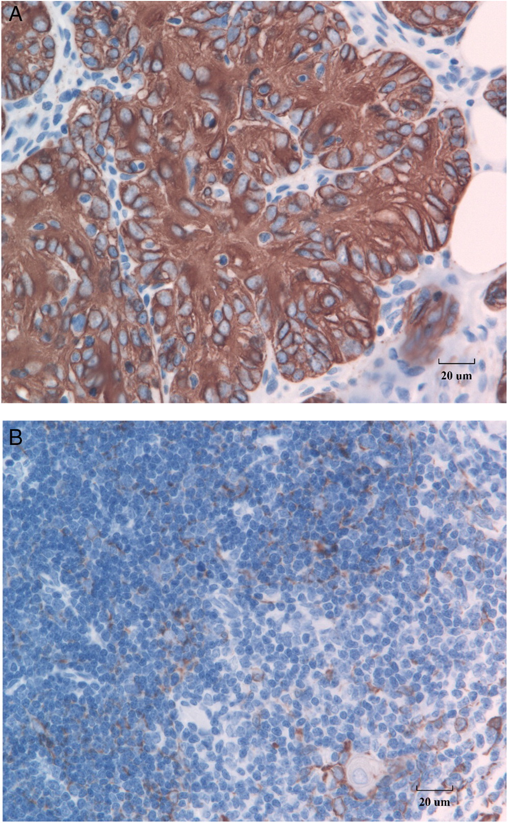
Figure 2B). Further, detection of cytokeratin in the SCID thymus reveals CK18 expression in the medullary spindle cell component of the thymic epithelial cells as well as cortical surface staining (Figure 3A), as compared with CK18 staining of normal thymic tissue control (
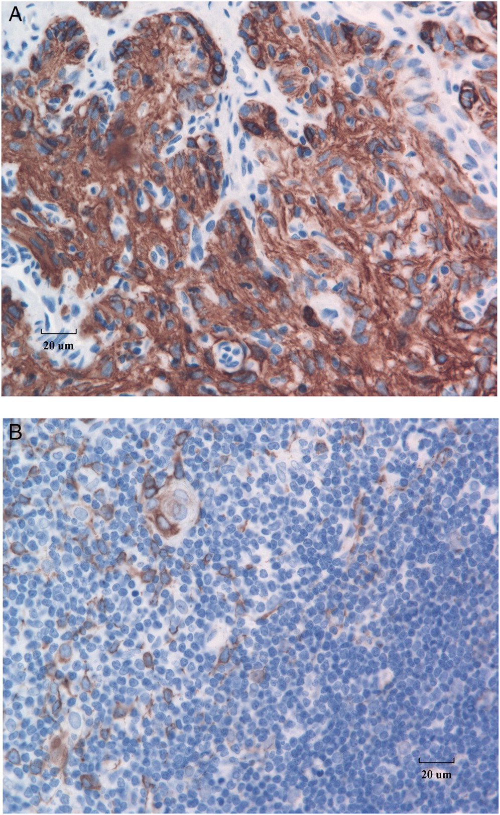
Figure 3B). Immunostaining with a cocktail of antibodies for CK8 and CK18 shows strong expression in both cortical polygonal thymic epithelial cells and spindle cells (Figure 4A) as compared with the staining pattern in a normal thymus (Figure 4B). In contrast to the healthy control thymus, in which cortical thymic epithelial cells stain negatively for CK5 and CK6 and medullary thymic epithelial cells stain positively for these markers (Figure 5B), in SCID, these cells all strongly express CK5 and CK6 (Figure 5A).
Figure 2:
Figure 2: Thymic dysplasia in SCID. (A) H&E stained thymus shows characteristic hypoplasia, loss of architecture, depletion of lymphocytes, loss of cortico-medullary differentiation, and absence of Hassall’s corpuscles. The thymic epithelial cells are reminiscent of primitive embryonic thymus. Clusters of micro-lobules of polygonal thymic epithelial cells are present, and towards the deeper areas, the epithelial cells assume spindle cell shapes. (B) Immunostaining for CD3, a pan T cell marker, reveals depletion of lymphocytes. (40× magnification)
Figure 3:
Figure 3: CK18 cytokeratin immunostaining of the SCID thymus. (A) Thymic epithelial cells in the cortical surfaces stain weakly for CK18, while the medullary spindle cell components express more CK18. (B) In the healthy control thymus only medullary thymic epithelial cells express CK18. (40× magnification)
Figure 4:
Figure 4: CK8 and CK18 expression in the SCID thymus. (A) Both cortical polygonal thymic epithelial cells as well as the spindle cells in the deeper regions have strong expression of both CK8 and CK18. (B) In the control thymus, cortical thymic epithelial cells stain positively for CK8, whereas CK18 is expressed by medullary thymic epithelial cells. (40× magnification)
Figure 5:
Figure 5: Expression of CK5 and CK6 in the SCID thymus. (A) SCID thymic epithelial cells all show strong expression for CK5 and CK6. (B) In contrast, the control thymus shows cortical thymic epithelial cells stain negatively for these markers, while medullary thymic epithelial cells stain positively. (40× magnification)
Three patients had all the hallmarks of a dysplastic thymus including loss, or near loss of lobular structure and cortico-medullary distinction, replacement of thymocytes with epithelioid cells and lack of Hassall’s corpuscles. In these patients the results supported the decision to perform BMT. The other 6 patients had various degrees of thymus gland abnormalities. In 5, Hassall’s corpuscles were either absent or barely developed suggestive of a profound T cell defect. Indeed, all 5 were offered BMT. One of these patients also had bone marrow failure (p3) which was by itself an indication for BMT. In 1 patient (p3) the thymus biopsy was critical in defining the final diagnosis of a novel type of SCID, CD3δ deficiency (Dadi et al. 2003), in another (p4) changes were mild to moderate and did not warrant further intervention.

Discussion

This report reviews the surgical approach and findings of transcervical thymus biopsy in children with a range of ages from infancy to adolescence. The thymus can be approached from various surgical routes. The traditional thymectomy involves a median sternotomy with its inherent risks and complications. The transcervical approach to the thymus is feasible in children. Identification of the lower part of the thyroid and often the thyrothymic ligament was helpful in identification of the thymus. As demonstrated in our series, the procedure can be performed in very young and severely ill children with minimal morbidity and a very low complication rate.
The diagnosis of an underlying congenital immunodeficiency may require the biopsy of lymphoid organs such as the thymus and lymph nodes. SCID as well as other forms of combined immunodeficiency are a group of fatal disorders unless treated with BMT. Typical cases of SCID present at infancy with failure to thrive, chronic diarrhea, Pneumocystis carinii pneumonia and oral thrush. Laboratory investigations frequently show profound lymphopenia.
In the past 15 years, we have increasingly recognized various types of profound T cell immunodeficiency that present in an atypical fashion. This may be caused by hypomorphic mutations in genes associated with SCID such as IL-2Rγ (Sharfe et al. 1997b; Somech and Roifman 2005) or in disorders caused by mutations in the IL-2Rα gene (Sharfe et al. 1997a) or Zap-70 (Roifman et al. 1989; Arpaia et al. 1994). In some of these cases, or others with an unknown genotype, the diagnosis of profound immunodeficiency may pose a challenge and hence the need for a thymic biopsy. We show here the importance of obtaining this tissue to make a sound decision concerning a high risk therapeutic procedure such as BMT.

Conclusions

This case series demonstrates that both positive and negative findings from a thymic biopsy contribute to the management of immunodeficient patients. Two patients were spared a BMT while another 8 patients were offered this life saving procedure. In addition, biopsies of the thymus have helped define new entities of primary immunodeficiency. The transcervical approach to the thymus can be performed with minimal morbidity.

REFERENCES

Arpaia E., Shahar M., Dadi H., Cohen A., and Roifman C.M. 1994. Defective T cell receptor signaling and CD8+ thymic selection in humans lacking Zap-70 kinase. Cell. 76(5):947–958.
Dadi H.K., Simon A.J., and Roifman C.M. 2003. Effect of CD3δ deficiency on maturation of α/β and γ/δ T-cell lineages in severe combined immunodeficiency. N. Engl. J. Med. 349(19):1821–1828.
Hong R. and Pellett J.W. 1978. Transcervical thymic biopsy in children with immunodeficiency. J. Pediatr. Surg. 13(4):427–428.
Janossy G., Thomas J.A., Bollum F.J., Granger S., Pizzolo G., Bradstock K.F., Wong L., McMichael A., Ganeshaguru K., and Hoffbrand A.V. 1980. The human thymic microenvironment: An immunohistologic study. J. Immunol. 125(1):202–212.
Joshi V.V., Oleske J.M., Saad S., Gadol C., Connor E., Bobila R., and Minnefor A.B. 1986. Thymus biopsy in children with acquired immunodeficiency syndrome. Arch. Pathol. Lab. Med. 110(9):837–842.
Leonard A.S., Mullholland M.W., and Filipovich A.H. 1985. Surgery of the immunodeficient child. Surg. Clin. North. Am. 65(6):1505–1525.
Roifman C.M. 2005. Studies of patients’ thymi aid in the discovery and characterization of immunodeficiency in humans. Immunol. Rev. 203:143–155.
Roifman C.M., Hummel D., Martinez-Valdez H., Thorner P., Doherty P.J., Pan S., Cohen F., and Cohen A. 1989. Depletion of CD8+ cells in human thymic medulla results in selective immune deficiency. J. Exp. Med. 170(6):2177–2182.
Sharfe N., Dadi H.K., Shahar M., and Roifman C.M. 1997a. Human immune disorder arising from mutation of the α chain of the interleukin-2 receptor. Proc. Natl. Acad. Sci. USA. 94(7):3168–3171.
Sharfe N., Shahar M., and Roifman C.M. 1997b. An interleukin-2 receptor gamma chain mutation with normal thymus morphology. J. Clin. Invest. 100(12):3036–3043.
Somech R. and Roifman C.M. 2005. Mutation analysis should be performed to rule out γc deficiency in children with functional severe combined immune deficiency despite apparently normal immunologic tests. J. Pediatr. 147(4):555–557.

Information & Authors

Information

Published In

cover image LymphoSign Journal
LymphoSign Journal
Volume 6Number 4December 2019
Pages: 141 - 147

History

Received: 12 September 2019
Accepted: 2 October 2019
Accepted manuscript online: 4 November 2019

Authors

Affiliations

Paolo Campisi [email protected]
Department of Otolaryngology—Head & Neck Surgery, Hospital for Sick Children, University of Toronto, Toronto, ON
Linda Vong
Division of Immunology and Allergy, Hospital for Sick Children, University of Toronto, Toronto, ON
Jonathan M. Sgro
Department of Otolaryngology—Head & Neck Surgery, Hospital for Sick Children, University of Toronto, Toronto, ON
Nikolaus Wolter
Department of Otolaryngology—Head & Neck Surgery, Hospital for Sick Children, University of Toronto, Toronto, ON
Bo Ngan
Department of Laboratory Medicine and Pathobiology, Hospital for Sick Children, University of Toronto, Toronto, ON
Jacob Friedberg
Department of Otolaryngology—Head & Neck Surgery, Hospital for Sick Children, University of Toronto, Toronto, ON

Funding Information

None

Metrics & Citations

Metrics

Other Metrics

Citations

Cite As

Export Citations

If you have the appropriate software installed, you can download article citation data to the citation manager of your choice. Simply select your manager software from the list below and click Download.

There are no citations for this item

View Options

View options

PDF

View PDF

Get Access

Login options

Check if you access through your login credentials or your institution to get full access on this article.

Subscribe

Click on the button below to subscribe to LymphoSign Journal

Purchase options

Purchase this article to get full access to it.

Restore your content access

Enter your email address to restore your content access:

Note: This functionality works only for purchases done as a guest. If you already have an account, log in to access the content to which you are entitled.

Media

Media

Other

Tables

Share Options

Share

Share the article link

Share on social media