Open access

Novel heterozygous NFKB1 mutation—variable penetrance in a family cohort

Publication: LymphoSign Journal
20 August 2019

Abstract

Background: Common variable immunodeficiency (CVID) is a term used to define a heterogeneous group of patients who commonly have hypogammaglobulinemia and variable degrees of modest T cell dysfunction. Recent advances made in next generation sequencing technologies have accelerated the identification of CVID disease-causing genes, including NFKB1, a component of the nuclear factor kappa-light-chain-enhancer of activated B cells (NF-κB) pathway.
Objective: We sought to identify the genetic defect in a 3-generation family of patients with CVID who presented with cytopenias, eczema, and recurrent sinopulmonary infections.
Methods: Whole exome sequencing and Sanger confirmation was performed, and a combination of molecular and cellular techniques used to assess the variant impact on B and T cell function.
Results: A novel heterozygous frameshift mutation in NFKB1, encoding the transcriptional regulator protein p50/p105, was identified. This resulted in c.1584dupG (p.M528fs). We demonstrate that c.1584dupG is a loss-of-function variant, responsible for reduced p105/p50 protein expression in affected individuals as well as variable increased CD21low B cell numbers.
Conclusion: This novel mutation affecting NFKB1 causes a CVID phenotype with variable clinical manifestations. Given the wide spectrum of age in this kindred, this may reflect diversity of phenotype expression, or more probably, age-related expression of typical features.
Statement of novelty: We report here a novel loss-of-function frameshift mutation in NFKB1.

Background

The classical response to invading pathogens involves physiological activation of the inflammatory response. Many of the individual, distinct, and complex immunological host defense pathways that make up this response meet at the level of nuclear factor kappa-light-chain-enhancer of activated B cells (NF-κB) activation, resulting in DNA binding and transcription (or transcriptional regulation) of several key genes. Thus, mutations in genes involved in the canonical and non-canonical NF-κB transcription factor pathways can broadly affect immune function, and have been shown to cause immunodeficiency disorders (Maffucci et al. 2016; Fliegauf and Grimbacher 2018).
NFKB1 encodes p50/p105 (NFKB1), which upon activation undergoes processing through phosphorylation and polyubiquitination of the Ankyrin repeat domain (ANK), and is cleaved into p50. The p50 transcription factor can homodimerize, leading to suppression of gene expression, or heterodimerize with the NF-κB class 2 subunits RelA (p65) or c-Rel, triggering gene expression (Lin et al. 2000; Salmerón et al. 2001; Karin and Lin 2002; Bonizzi and Karin 2004; Vallabhapurapu and Karin 2009).
Activation of the canonical NFKB1 pathway by a variety of receptors, including interleukin (IL)-1R, tumor necrosis factor receptor (TNFR), toll-like receptor (TLR), CD40, CD30, RANK, T cell receptor (TCR) and B cell receptor (BCR), mediated by the IĸBα and IKK complex, ultimately results in p50/RelA-dependant gene expression. Given the important role of NFKB1 in the cell regulation, aberrations affecting any component of this cascade are likely to have detrimental effects on host defense.
Recently, mutations affecting NFKB1 were reported in individuals diagnosed with common variable immunodeficiency (CVID), characterized by hypogammaglobulinemia and modest T cell dysfunction. Results of clinical and immune laboratory presentation varied widely between those affected, however, all resulted from perturbed p50/p105 expression and subsequent dysfunction of NFKB1 pathway activation and gene transcription (Fliegauf et al. 2015; Boztug et al. 2016; Kaustio et al. 2017; Lougaris et al. 2017; Tuijnenburg et al. 2018).
In this case series, we describe a 3-generation Irish-Canadian family with autosomal dominant CVID and include details of clinical and immune laboratory phenotype as well as genetic evaluation. Results of whole exome sequencing (WES) revealed a novel heterozygous frameshift mutation in NFKB1 resulting in loss-of-function.

Methods

Patients

All patient data and samples were obtained in accordance with the Declaration of Helsinki and the Research Ethics Board at The Hospital for Sick Children. Patient data was compiled prospectively and retrospectively from medical records and entered into the Primary Immunodeficiency Registry and Tissue Bank (REB protocol No. 1000005598). Informed consent was obtained for genetic testing, including WES, as well as extensive investigations to unravel the functional effect of immune abnormalities.

DNA isolation and preparation

Genomic DNA was isolated and extracted from peripheral blood lymphocytes (PBL) using the Geneaid genomic DNA extraction kit (Geneaid Mini Kit, ON, Canada), in accordance with the manufacturer’s instructions.

Targeted exome sequencing and data analysis

DNA was quantified using the Qubit DNA HS Assay (Life Technologies, Carlsbad, CA, USA) and 100 ng of input DNA subsequently used for library preparation (Ion AmpliSeq Exome Kit; Life Technologies). The exome library was immobilized on Ion PI™ Ion Sphere particles (Ion PI Template OT2 200 Kit v3; Life Technologies). Sequencing was performed utilizing the Ion PI™ Sequencing 200 Kit v3 and Ion PI™ Chip v2 in the Ion Proton semiconductor sequencing system, according to the manufacturer’s recommendations (Life Technologies).

Coverage and variant validation

Alignment and variant calling were performed with Torrent Suite (v4.0) on the Ion Proton Server using the Ion Proton AmpliSeq germline low-stringency setting and the hg19 reference genome. The variants were annotated using an in-house annotation pipeline 35 (based on Annovar; November 2014 version), 36 and RefSeq gene models (downloaded from UCSC on 1 August 2015).

Flow cytometry-based immunophenotyping and T cell proliferation assays

B cell phenotypes were determined by flow cytometric analysis using the 4-laser BD analyzer LSR II CFI, BGRV (BD Biosciences, San Jose, CA, USA). Antibodies used included the following: CD19-BV711, CD27-BV421, IgD-PE-Cy7, CD24-BB515, CD38-allophycocyanin, CD21-PE-CF594, CD45RO-PE, CD197-BV421, and CD196-PE-Cy7 (BD Biosciences, San Jose, CA, USA).
Lymphocyte proliferative responses to mitogens, including PHA and the combination of anti-CD3 and anti-CD28 antibodies, and to a panel of recall antigens, including Candida species, tetanus toxoid, herpes zoster, and cytomegalovirus, were determined by H-thymidine incorporation (Arpaia et al. 1994). All assays were performed in triplicate and compared with simultaneously stimulated cells from healthy control subjects.

Western blot analysis

PBL was collected from patients and healthy controls, and T lymphocytes selectively cultured (CD3/CD28 Human T activator Dynabeads; Gibco, Life Technologies, Carlsbad, CA, USA) in the presence of IL-2. Cells were lysed on ice using lysis buffer (1% Triton X-Vanadate) and loaded onto 12% polyacrylamide gel for size fractionation (NuPage 4%–12% Bis-Tris Gel, Invitrogen, Rockford, IL, USA). Proteins were transferred onto polyvinylidene fluoride membrane in accordance with standard Western blotting protocols. p105 and p50 proteins were detected using rabbit anti-NF-κB1 p105/p50 antibody (#3035 Cell Signaling, Danvers, MA, USA; 1:1000 dilution). Anti-rabbit secondary antibody coupled to horseradish peroxidase (7074S, Cell Signaling; Danvers, MA, USA; 1:5000 dilution) was used for signal detection. Immunoreactive bands were visualized with ECL Western Blotting Substrate (ThermoScientific, Life Technologies, Carlsbad, CA, USA).

Serum concentration of immunoglobulins and specific antibodies

Serum concentrations of immunoglobulins (IgG, IgA, and IgM) were measured using nephelometry, and IgE concentrations were measured by using radioimmunoassay with the IgE PRIST kit (Pharmacia Diagnostics, Dorval, QC, Canada). Levels of serum tetanus toxoid and pneumococcus (pneumococcal capsular polysaccharide) were measured by means of ELISA, according to the manufacturer’s instructions (Binding Site, Birmingham, UK). Serum antibodies to measles, mumps, and rubella were measured by ELISA with kits available from Euroimmune (Gross-Groenau, Germany). Serum isohemagglutinin titers were determined by means of 2-fold serial dilution with erythrocytes and are reported as the antiglobulin phase, the dilution at which macroscopic agglutination is observed.

Case report

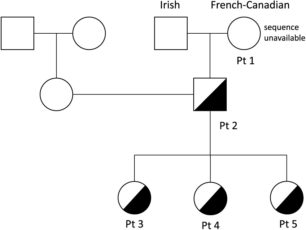
A pedigree of the family members is shown in Figure 1.
Figure 1:
Figure 1: Pedigree of kindred with heterozygous NFKB1 mutations. DNA sample from patient 1 (Pt 1) was unavailable for sequencing. Patient 2 (Pt 2), son of patient 1, as well as patients 3, 4, and 5 (Pt 3, 4, 5) carry the heterozygous NFKB1 c.1584dupG, p.M528fs mutation.

Patient 1

Patient 1 was a product of non-consanguineous parents of Irish and French descent. She presented at young adulthood, age 23 years, with recurrent prolonged upper respiratory tract infections (10/year), chest X-ray-proven pneumonias (5/year), chronic sinusitis, and recurrent otitis with purulent otorrhea, fatigue, and weight loss. Her childhood clinical history was unremarkable, with uneventful chickenpox, measles, and mumps infections. She had 1 episode of arthritis at age 2 years and normal echocardiography on examination. A tonsillectomy was performed at 5 years of age.
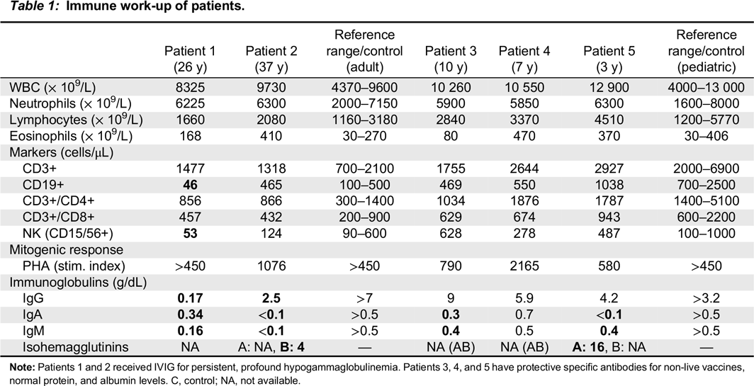
During her initial evaluation with our Immunology team at age 26 years, she presented with cylindrical bronchiectasis on bronchoscopy of the left lower lobe and the lingula. She had normal tuberculin test results and sweat chloride levels (7 mEq/L). Immunological evaluation (Table 1) showed pan-hypogammaglobulinemia, with low IgG, IgM, and IgA levels, as well as low anti-tetanus toxoid titer (0.03 ng/mL). IgE levels were normal, as were protein and albumin levels. Her CBC and differential was unremarkable. Lymphocyte counts were normal, however, B cell counts were almost absent with only 46 cells/μL of CD19+ cells alongside decreased NK cell numbers of 53 cells/μL. Assay for spontaneous suppressive cell activity assay was positive, and E-rosette and AET rosette assessment of T lymphocytes were normal at 58% and 70%, respectively. Results of mixed lymphocyte reaction were also normal. Lymphocyte proliferation responses to all mitogens except STA were normal, however, antigen stimulation responses to Candida and Tuberculin PPD were decreased.
Table 1:
Table 1: Immune work-up of patients.

Note: Patients 1 and 2 received IVIG for persistent, profound hypogammaglobulinemia. Patients 3, 4, and 5 have protective specific antibodies for non-live vaccines, normal protein, and albumin levels. C, control; NA, not available.

Due to her clinical presentation and diminished immune responses, she was started on fresh frozen plasma (FFP), the only IgG replacement option that was available at the time she was diagnosed. She was eventually transferred to intramuscular and then intravenous immunoglobulin therapy. Laboratory assessments showed adequate IgG replacement. Unfortunately, the patient contracted blood product-related Hepatitis C infection resulting in liver cirrhosis, portal hypertension, massive ascites and varicose veins requiring multiply ligation and sclerotherapy due to bleeding, and eventually liver failure. Other immunodeficiency related clinical manifestations include chronic cough, yellow sputum, Mycoplasma colonization, chronic Salmonella gastroenteritis which resolved after 6 months of antibiotic therapy, chronic non-infectious diarrhea, and hepatosplenomegaly.
At the age of 36 years, the patient developed ophthalmic melanoma resulting in loss of vision in the affected eye. She suffered severe bronchiectasis with pyocele 2 years later for which she underwent left lower lobe lobectomy. Concurrently, she slowly developed progressing cerebellar symptoms with abnormal Romberg and heel/toe gait, however, CT imaging of the head was unremarkable.
Following a prolonged course of weight loss, intermittent fevers, and 1 episode of septicaemia, worsening productive cough, failed empiric antibiotic therapy, and increased fatigue, the patient was subsequently evaluated for malignancy. She was diagnosed with lymphoma and later died at age 45 years.

Patient 2

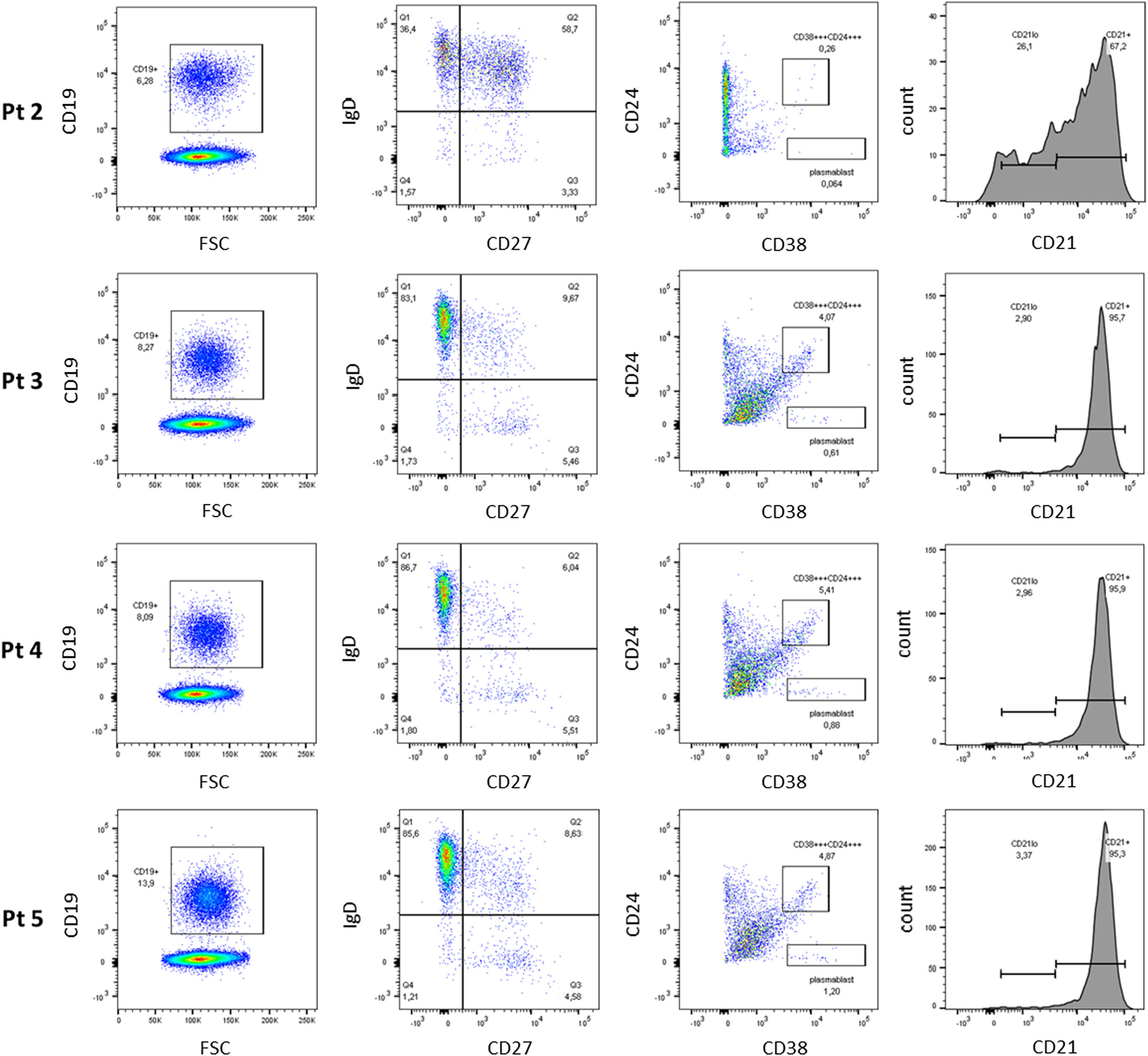
Patient 2, the son of patient 1, was evaluated at 1 year of age and found to have low IgG, IgM, and IgA levels (Table 1). At the age of 10 years he developed chronic immune thrombocytopenia and has been on regular monthly intravenous IgG replacement therapy since age 11 years. He was diagnosed with persistent abdominal lymphadenomegaly, and percutaneous needle aspiration biopsy showed follicular hyperplasia. His CBC and differential were unremarkable, with normal numbers of circulating CD3+, CD4+, CD8+, CD19+ and NK cells. Stimulation of T cells with anti-CD3 and anti-CD28 was markedly reduced, implying a T cell defect. In vitro lymphocyte responses to a series of antigens, including Candida species, tetanus toxoid, zoster, and cytomegalovirus, were also extremely low indicating the antigen specific memory T cells were either lacking or present at low frequencies (Figure 2). Assessment of B cell phenotype by flow cytometry showed increased number of IgD+ memory cells, but diminished class switching (CD38+/CD24+) (Figure 3). No transitional B cells and elevated number in CD21low B cells were detected.
Figure 2:
Figure 2: Reduced lymphocyte proliferation responses to antigens. T lymphocyte proliferation responses in patient 2 were reduced when assayed against a panel of recall antigens including Candida, tetanus toxoid, herpes zoster, and cytomegalovirus (CMV).
Figure 3:
Figure 3: Assessment of class-switched memory B cells. Flow cytometry analysis shows increased number of IgD+ memory B cells, but diminished class-switching (CD38+ CD24+) in Patient 2 (Pt 2). No transitional B cells and increased CD21low and CD27+ IgD+ B cell counts were found. Assessment of B cell phenotype reveals normal age appropriate populations in patients 3, 4, and 5.

Patient 3

Patient 3, daughter of patient 2, is a 10 year old with a history of 3 episodes of chickenpox at ages 3, 5, and 7 years. Given the clinical history of her grandmother and father, live viral vaccines were avoided. Her CBC and differential were unremarkable, with normal B and T cell lymphocyte function. She had normal IgG levels with adequate specific antibody titers, but decreased levels of IgA and IgM. Her varicella zoster virus specific IgG antibody titers were negative. Despite 3 episodes of chickenpox, she was unable to mount an appropriate immune response.
Patient 4

Patient 4, daughter of patient 2, is a 7 year old female. She too suffered 3 episodes of chickenpox. Her immunological evaluations to date have been unremarkable. Like her sister, her varicella zoster virus specific antibody titers were negative.

Patient 5

Patient 5, the youngest sibling of patients 3 and 4, is a 3 year old female with eczema and repeated upper respiratory tract infection. While her T cell response to phytohemagglutinin was normal, the patient’s response to mitogen stimulation with anti-CD3 were decreased. She also had decreased IgA and IgM but normal IgG levels with adequate antibody responses to vaccination.

Identification of NFKB1 mutation

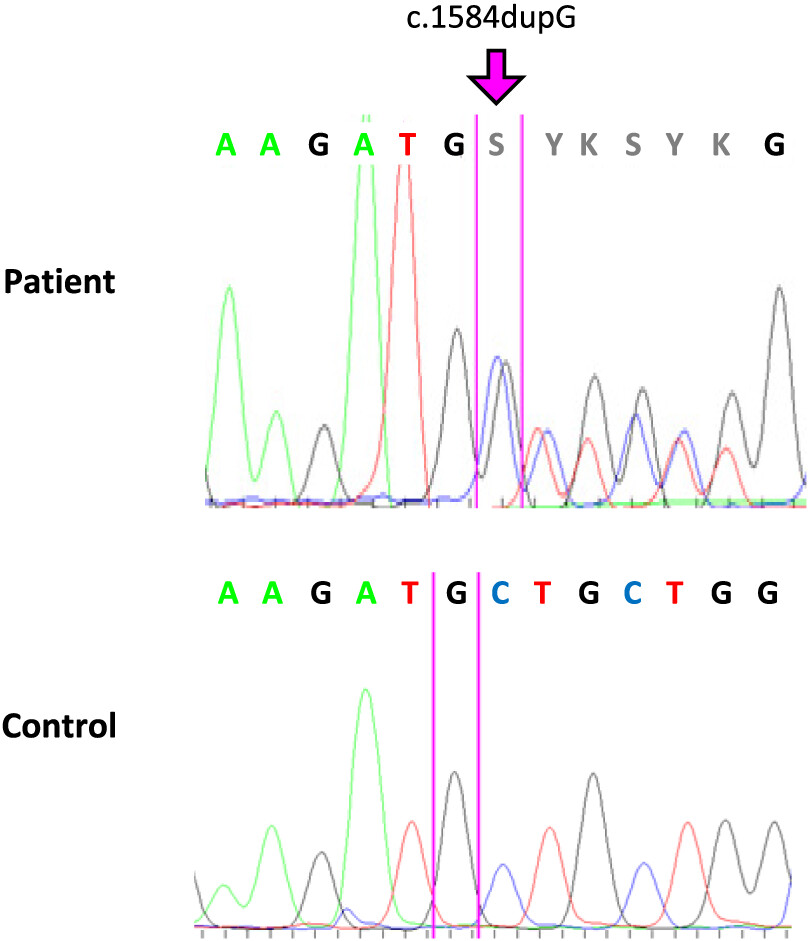
WES was performed on patient 2. We identified a novel heterozygous variant in the gene NFKB1 (NM_03998.3), c.1584dupG, resulting in p.M528fs (Figures 4 and 5). This was confirmed by Sanger sequencing of patients 2–5 and segregated with disease. The M528fs mutation in NFKB1 has not been previously reported in dbSNP, ClinVar, or HGMD. Due to unavailability of genetic material, presence of the NFKB1 variant in patient 1 could not be confirmed.
Figure 4:
Figure 4: Heterozygous mutation in NFKB1 in patients with CVID. Electropherogram of the patient sequence (upper panel) shows the heterozygous mutation c.1584dupG, resulting in the frameshift p.M528fs. The wild-type control sequence is shown in the lower panel.
Figure 5:
Figure 5: Mutation in NFKB1 is localized upstream of the ankyrin repeat domain. NFKB1 is composed of 4 distinct domains; Rel homology domain (RHD), glycine rich region (GRR), ankyrin repeat domain (ANK), and death domain (DD). Following phosphorylation and polyubiquitination, the p105 protein is cleaved at amino acid 433 to generate p50. The frameshift mutation identified in our patients, p.M528fs, likely leads to protein instability and increased degradation.

Signalling evaluation

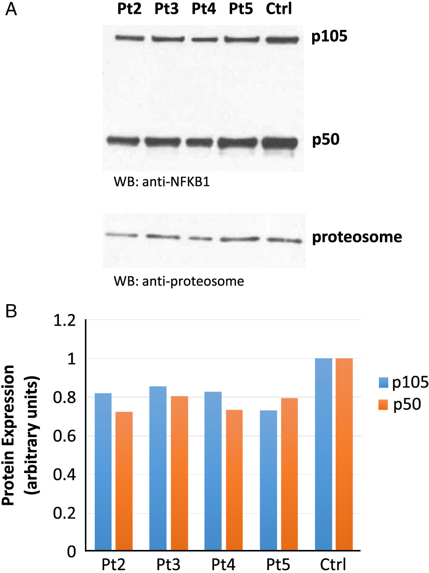
Western blot evaluation of samples obtained from our patients carrying the novel heterozygous NFKB1 mutation showed a marked decrease in p50 and p105 protein levels, indicating that this variant is a loss-of-function mutation (Figure 6).
Figure 6:
Figure 6: Western blot analysis of NFKB1 expression. (A) Detection of p50/p105 protein levels in patients with the heterozygous p.M528fs mutation compared to control. (B) Densitometric analysis reveals reduced levels of the precursor protein p105 as well as the p50 subunit in patients 2–5. Results are normalized against α-proteosome expression to ensure equal loading.

Discussion

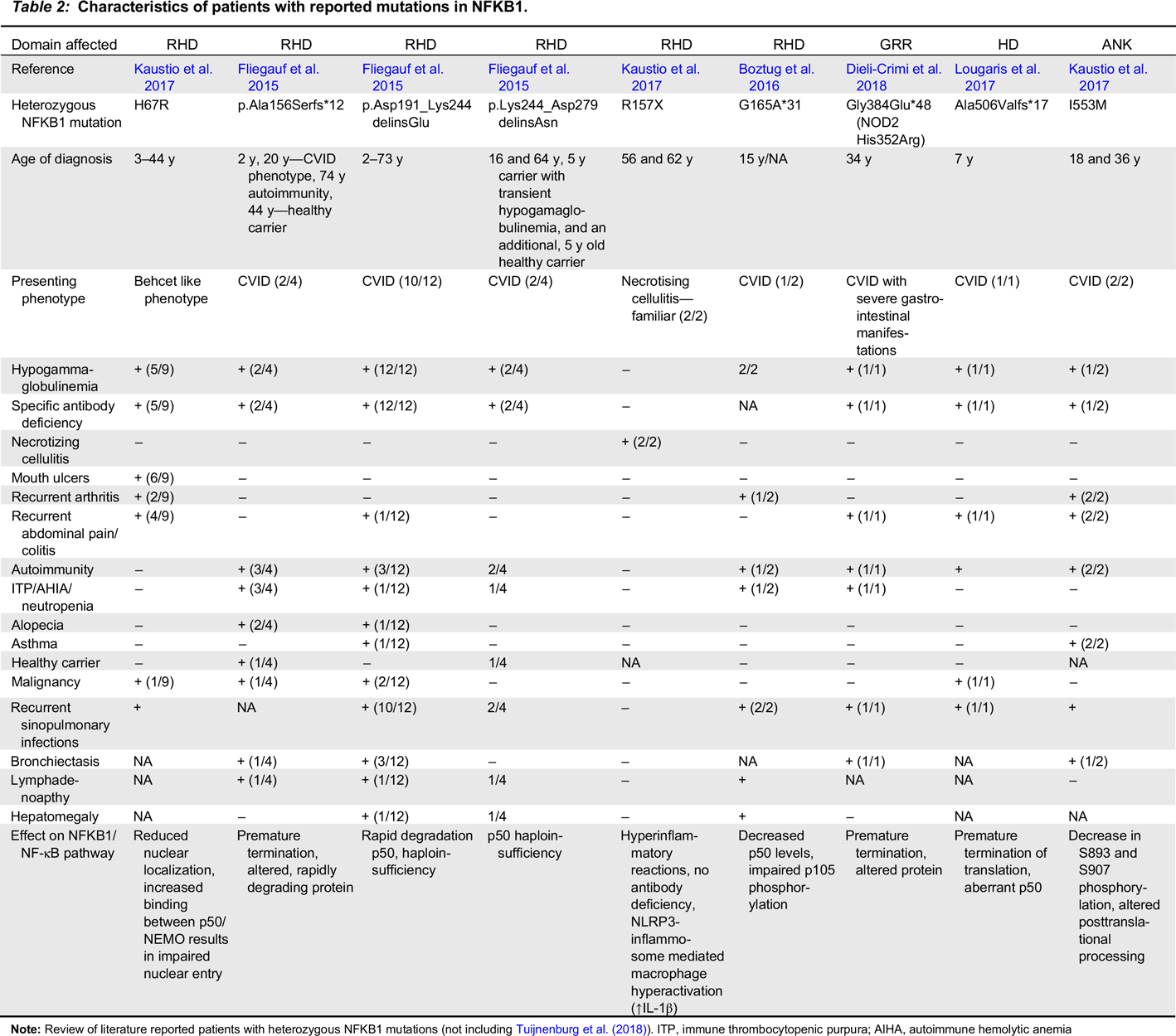
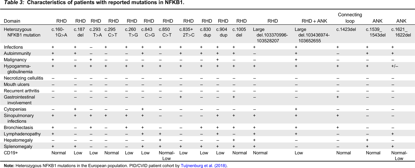
The NFKB1 gene encodes p50/p105, which upon receptor activation undergoes proteosomal processing of the C-terminal to produce p50, a transcription factor that forms, among others, a p50/RelA heterodimer to regulate gene transcription. Heterozygous NFKB1 mutations are described in the literature with variable clinical and laboratory manifestations (summarized in Tables 2 and 3). The features in these patients vary even within families. The most common features associated with heterozygous NFKB1 mutations are hypogammaglobulinemia associated with recurrent sinopulmonary infections, as well as autoimmune disorders including immune mediated cytopenias and enteropathy. Some patients suffered benign or malignant lymphoproliferation (Tuijnenburg et al. 2018). In addition, other phenotypes lacking infections, such as necrotizing cellulitis, were associated with distinct mutations in NFKB1 (Kaustio et al. 2017).
Table 2:
Table 2: Characteristics of patients with reported mutations in NFKB1.

Note: Review of literature reported patients with heterozygous NFKB1 mutations (not including Tuijnenburg et al. (2018)). ITP, immune thrombocytopenic purpura; AIHA, autoimmune hemolytic anemia

Table 3:
Table 3: Characteristics of patients with reported mutations in NFKB1.

Note: Heterozygous NFKB1 mutations in the European population. PID/CVID patient cohort by Tuijnenburg et al. (2018).

Within our kindred, analysis of B cell populations demonstrated a reduction in class-switched memory B cells and accumulation of CD21low B cells, with a broad range of non-switched memory B cells, and an inability to generate plasma blasts (CD38+/CD27++). The only difference between asymptomatic and clinically affected NFKB1 variant carriers was the increased numbers of CD21low B cell population. However, it is plausible to speculate that changes in B cell subpopulations, specific antibody titres, as well as clinical susceptibility to infections and autoimmunity are age-related.
p50/p105 protein levels were consistently reduced by approximately 40% in symptomatic individuals with CVID (Tuijnenburg et al. 2018). The T cell phenotypic presentation and function was generally unaffected, however decreased NK cell numbers were observed (Dieli-Crimi et al. 2018; Tuijnenburg et al. 2018). T cell proliferation against anti-CD3/anti-CD28 in patients of a European cohort were previously shown to be intact (Tuijnenburg et al. 2018). In our cohort, patient 1 and patient 4 had aberrant T cell proliferation responses to the mitogen CD3.

Conclusion

Mutations that disrupt the function of p105/p50 lead to defects in the canonical NF-κB pathway, resulting in autosomal dominant CVID with highly variable features, even within 1 family. Our kindred represents a perfect example of this diversity, from complete antibody deficiency and malignancy to subtle immune aberrations and clinical manifestations which are predominately age-related. As CVID is known to be predominately an immunodeficiency affecting adults, it is reasonable to speculate that the children in our kindred display, at this stage, only the early signs of CVID.

Abbreviations used

NFKB1
nuclear factor kappa-light-chain-enhancer 1 of activated B cells
NF-κB
nuclear factor kappa-light-chain-enhancer of activated B cells
CVID
common variable immunodeficiency
ANK
ankyrin repeat domain
AD-EDA-ID/NEMO
autosomal dominant ectodermal dysplasia with immune deficiency
DD
death domain
NLRP3
NLR family pyrin domain containing 3
RHD
Rel homology domain
PMA
Phorbol 12-myristate 13-acetate
SCID
severe combined immunodeficiency
TCR
T cell receptor
TCRvB
T cell receptor v Beta
WES
whole exome sequencing

REFERENCES

Arpaia E., Shahar M., Dadi H., Cohen A., and Roifman C.M. 1994. Defective T cell receptor signaling and CD8+ thymic selection in humans lacking Zap-70 kinase. Cell. 76(5):947–958.
Bonizzi G. and Karin M. 2004. The two NF-κB activation pathways and their role in innate and adaptive immunity. Trends Immunol. 25(6):280–288.
Boztug H., Hirschmugl T., Holter W., Lakatos K., Kager L., Trapin D., Pickl W., Förster-Waldl E., and Boztug K. 2016. NF-κB1 haploinsufficiency causing immunodeficiency and EBV-driven lymphoproliferation. J. Clin. Immunol. 36:533–540.
Dieli-Crimi R., Martínez-Gallo M., Franco-Jarava C., Antolin M., Blasco L., Paramonov I., Semidey M.E., Álvarez Fernández A., Molero X., Velásquez J., Martín-Nalda A., Pujol-Borrell R., and Colobran R. 2018. Th1-skewed profile and excessive production of proinflammatory cytokines in a NFKB1-deficient patient with CVID and severe gastrointestinal manifestations. Clin. Immunol. 195:49–58.
Fliegauf M. and Grimbacher B. 2018. Nuclear factor κB mutations in human subjects: The devil is in the details. J. Allergy Clin. Immunol. 142(4):1062–1065.
Fliegauf M., Bryant V.L., Frede N., Slade C., Woon S.T., Lehnert K., Winzer S., Bulashevska A., Scerri T., Leung E., Jordan A., Keller B., de Vries E., Cao H., Yang F., Schäffer A.A., Warnatz K., Browett P., Douglass J., Ameratunga R.V., van der Meer J.W., and Grimbacher B. 2015. Haploinsufficiency of the NF-κB1 subunit p50 in common variable immunodeficiency. Am. J. Hum. Genet. 97:389–403.
Karin M. and Lin A. 2002. NF-κB at the crossroads of life and death. Nat. Immunol. 3(3):221–227.
Kaustio M., Haapaniemi E., Göös H., Hautala T., Park G., Syrjänen J., Einarsdottir E., Sahu B., Kilpinen S., Rounioja S., Fogarty C.L., Glumoff V., Kulmala P., Katayama S., Tamene F., Trotta L., Morgunova E., Krjutškov K., Nurmi K., Eklund K., Lagerstedt A., Helminen M., Martelius T., Mustjoki S., Taipale J., Saarela J., Kere J., Varjosalo M., and Seppänen M. 2017. Damaging heterozygous mutations in NFKB1 lead to diverse immunologic phenotypes. J. Allergy Clin. Immunol. 140(3):782–796.
Lin L., DeMartino G.N., and Greene W.C. 2000. Cotranslational dimerization of the Rel homology domain of NF-κB1 generates p50–p105 heterodimers and is required for effective p50 production. EMBO J. 19(17):4712–4722.
Lougaris V., Moratto D., Baronio M., Tampella G., van der Meer J.W.M., Badolato R., Fliegauf M., and Plebani A. 2017. Early and late B-cell developmental impairment in nuclear factor kappa B, subunit 1-mutated common variable immunodeficiency disease. J. Allergy Clin. Immunol. 139(1):349–352.e1.
Maffucci P., Filion C.A., Boisson B., Itan Y., Shang L., Casanova J.-L., and Cunningham-Rundles C. 2016. Genetic diagnosis using whole exome sequencing in common variable immunodeficiency. Front. Immunol. 7:220.
Salmerón A., Janzen J., Soneji Y., Bump N., Kamens J., Allen H., and Ley S.C. 2001. Direct phosphorylation of NF-κB1 p105 by the IκB kinase complex on serine 927 is essential for signal-induced p105 proteolysis. J. Biol. Chem. 276(25):22215–22222.
Tuijnenburg P., Lango Allen H., Burns S.O., Greene D., Jansen M.H., Staples E., Stephens J., Carss K.J., Biasci D., Baxendale H., Thomas M., Chandra A., Kiani-Alikhan S., Longhurst H.J., Seneviratne S.L., Oksenhendler E., Simeoni I., de Bree G.J., Tool A.T.J., van Leeuwen E.M.M., Ebberink E.H.T.M., Meijer A.B., Tuna S., Whitehorn D., Brown M., Turro E., Thrasher A.J., Smith K.G.C., Thaventhiran J.E., Kuijpers T.W., and NIHR BioResource-Rare Diseases Consortium. 2018. Loss-of-function nuclear factor κB subunit 1 (NFKB1) variants are the most common monogenic cause of common variable immunodeficiency in Europeans. J. Allergy Clin. Immunol. 142(4):1285–1296.
Vallabhapurapu S. and Karin M. 2009. Regulation and function of NF-κB transcription factors in the immune system. Annu. Rev. Immunol. 27(1):693–733.

Information & Authors

Information

Published In

cover image LymphoSign Journal
LymphoSign Journal
Volume 6Number 3September 2019
Pages: 95 - 105

History

Received: 4 July 2019
Accepted: 8 August 2019
Accepted manuscript online: 20 August 2019

Authors

Affiliations

Amarilla B. Mandola [email protected]
The Division of Immunology and Allergy, Department of Pediatrics, Hospital for Sick Children and the University of Toronto, Toronto, ON
The Canadian Centre for Primary Immunodeficiency and the Jeffrey Modell Research Laboratory for the Diagnosis of Primary Immunodeficiency, Hospital for Sick Children, Toronto, ON
Daniele Merico
The Centre for Applied Genomics (TCAG), Program in Genetics and Genome Biology, The Hospital for Sick Children, Toronto, ON
Vevo Therapeutics, San Francisco, CA
Nigel Sharfe
The Division of Immunology and Allergy, Department of Pediatrics, Hospital for Sick Children and the University of Toronto, Toronto, ON
The Canadian Centre for Primary Immunodeficiency and the Jeffrey Modell Research Laboratory for the Diagnosis of Primary Immunodeficiency, Hospital for Sick Children, Toronto, ON

Metrics & Citations

Metrics

Other Metrics

Citations

Cite As

Export Citations

If you have the appropriate software installed, you can download article citation data to the citation manager of your choice. Simply select your manager software from the list below and click Download.

There are no citations for this item

View Options

View options

PDF

View PDF

Get Access

Login options

Check if you access through your login credentials or your institution to get full access on this article.

Subscribe

Click on the button below to subscribe to LymphoSign Journal

Purchase options

Purchase this article to get full access to it.

Restore your content access

Enter your email address to restore your content access:

Note: This functionality works only for purchases done as a guest. If you already have an account, log in to access the content to which you are entitled.

Media

Media

Other

Tables

Share Options

Share

Share the article link

Share on social media