The spectrum of multisystem inflammatory syndrome (MIS-C) in children infected with severe acute respiratory syndrome coronavirus 2
Abstract
Introduction: The impact of SARS-CoV-2 infections in children has generally been described as relatively benign. However, since April 2020, there have been reports of a new multisystem inflammatory illness affecting children and related to COVID-19 termed multisystem inflammatory syndrome in children (MIS-C).
Aim: To describe 3 cases of children diagnosed with MIS-C and discuss the disease spectrum.
Methods: We collected and reviewed data from 3 cases diagnosed with MIS-C admitted to our pediatric ward between October 2020 and January 2021.
Discussion: MIS-C is a newly described disease that spans a spectrum of phenotypes and severity, and while it shares clinical similarities with Kawasaki disease, it has a unique set of epidemiological, laboratory, and prognostic characteristics. In this review, we hope to add to the understanding of this new entity.
Statement of Novelty: This report discusses 3 cases of MIS-C and elaborates on the spectrum and immunology of this entity. Our cases are unique in their relatively wide spectrum and variability. We hope our own experience with MIS-C adds to the accumulating knowledge and understanding of this emerging disease.
Background
Coronavirus disease 2019 (COVID-19) is a rapidly spreading pandemic caused by the novel severe acute respiratory syndrome coronavirus 2 (SARS-CoV-2). Since its origins in Wuhan, Hubei Province of China, in December 2019, the disease has affected more than 100 million people worldwide, and with over 2 million deaths as of February 2021 according to the World Health Organization (WHO) COVID-19 Dashboard (Organisation 2021). As of March 2021, COVID-19 has resulted in over 6,000 deaths in Israel, while more than 500 patients are currently in a severe condition, including 250 whom are mechanically ventilated (Datadashboard.health.gov.il n.d).
The impact of SARS-CoV-2 infections in children has generally been described as relatively benign (Castagnoli et al. 2020). However, since April 2020, there have been reports of a new multisystem inflammatory illness affecting children, related to COVID-19, termed multisystem inflammatory syndrome in children (MIS-C), sometimes also described as “pediatric inflammatory multisystem syndrome temporally associated with SARS-CoV-2” (PIMS) (Ng et al. 2020; Riphagen et al. 2020; Verdoni et al. 2020; Viner and Whittaker 2020). Patients with MIS-C exhibit similar symptoms to those found in Kawasaki disease (KD), and streptococcal and staphylococcal toxic shock syndromes (TSS); however, there are several key clinical, epidemiological, and importantly immunological features that are unique to this syndrome (RCPCH n.d; Arad et al. 2011; Abrams et al. 2020; Ahmed et al. 2020; Consiglio et al. 2020). Here, we describe 3 cases of this newly described disease and discuss its spectrum.
Case presentations
All patients described below were admitted to our pediatric ward between October 2020 and January 2021. All clinical and laboratory manifestations are summarized in Table 1.
Table 1:
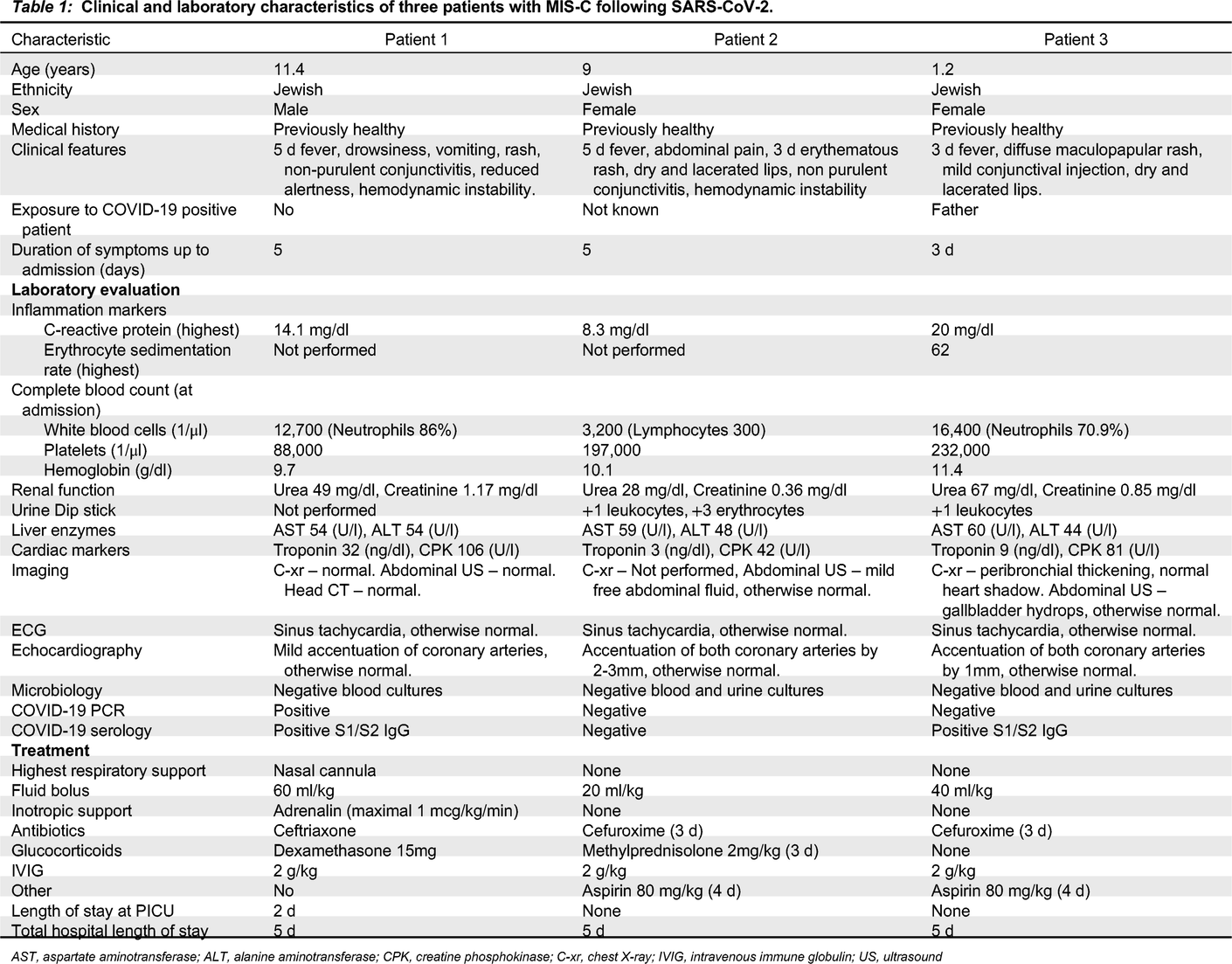
AST, aspartate aminotransferase; ALT, alanine aminotransferase; CPK, creatine phosphokinase; C-xr, chest X-ray; IVIG, intravenous immune globulin; US, ultrasound
Patient 1, an 11-year-old male, presented in the pediatric emergency room with a medical history of 5 days fever and vomiting, new onset of drowsiness, diffuse maculopapular rash, non–purulent conjunctivitis, and cracked red lips. On physical examination, he was found to have meningeal irritation and nuchal rigidity. He was treated with a fluid bolus and antibiotics on a working diagnosis of bacterial meningitis, but within an hour he deteriorated and developed hemodynamic instability leading to cardiovascular shock that required vasopressor support.
Patient 2, a 9-year-old female, presented with abdominal pain. She was suspected to have appendicitis based on the clinical picture, and was hospitalized for further investigation. While hospitalized, she developed KD-like symptoms including fever, rash, cracked lips, and non-purulent conjunctivitis. In addition, she also developed low blood pressure that responded well to fluid treatment.
Patient 3, a 1-year-old female, presented with a 3 day fever, and later developed a rash, mild conjunctival injection, and cracked lips.
All patients had strikingly elevated inflammation markers. Other hematological abnormalities are presented in Table 1. Two patients (1 and 3), had a transient acute kidney injury, while all 3 had a mild elevation of liver enzymes that later resolved. Patient 1 had a mildly elevated troponin I, while patient 2 had sterile pyuria on admission. Chest X-ray was performed for all patients with no significant findings. Patient 2 had low amounts of free abdominal fluid on ultrasonography; patients 3 had hydrops of the gallbladder. Electrocardiography was normal except for sinus tachycardia in all patients. Echocardiography demonstrated prominent coronary arteries for all patients, cardiac function was normal; patient 1 had a mild pericardial effusion.
All patients underwent extensive microbiological investigation (Table 1).
SARS-CoV-2 polymerase chain reaction (PCR) testing was positive for patient 1, while Anti-SARS-CoV-2 immunoglobulin G (IgG) was positive for patients 1 and 3. The serology testing was performed using the DiaSorin (Saluggia VC, Italy) Liaison SARS-CoV-2 S1/S2 IgG assay, which detects antibodies specific to the SARS-CoV-2 spike (S) proteins. All other infectious investigations were negative.
All patients were treated with a fluid bolus and intravenous immunoglobulin (IVIG). Treatment with wide spectrum antibiotics was initiated in all 3 patients until negative results of blood and urine cultures. Patients 1 and 2 were also treated with glucocorticoids. Patient 1 was admitted to the pediatric intensive care unit (PICU) for vasopressor (adrenaline) and respiratory support (nasal cannula) for 1 day, the patient responded well to treatment with a rapid clinical improvement and was discharged from the PICU to the pediatric ward after 2 days. All 3 patients were discharged home after 5 days.
Discussion
Accumulating evidence that an inflammatory syndrome may follow SARS-CoV-2 infection in some children is in contrast to the general impression that COVID-19 is mostly asymptomatic in children but may present with mild respiratory or gastrointestinal symptoms (Castagnoli et al. 2020).
The Royal College of Pediatrics and Child Health (RCPCH), center for disease control (CDC) and WHO have fairly similar but not identical criteria for the emerging condition of MIS-C. All 3 cite inflammation, and single or multi organ dysfunction, although the RCPCH does not require virological evidence, while the WHO and the CDC criteria include viral positive PCR or serology, or close contact to a known COVID-19 patient (RCPCH n.d.; www.who.int n.d.; Centers for Disease Control and Prevention 2021; Riphagen et al. 2020). All 3 cases in our report fulfilled the RCPCH case definition, while patients 1 and 3 also satisfy the CDC and WHO criteria. Patient 2 had no virological evidence of infection; however, the presentation is highly suggestive of the disease (RCPCH n.d; www.who.int n.d.; Centers for Disease Control and Prevention 2021).
MIS-C appears to span a spectrum; this is reflected in the patients we described. Most patients in case series and reviews were above 5 years of age (Ahmed et al. 2020; Ramcharan et al. 2020; Bustos et al. 2021); however, infants with the disease have also been described (Bautista-Rodriguez et al. 2021). The disease has also been reported across different races, ethnicities, and countries (Ahmed et al. 2020; Ramcharan et al. 2020; Bustos et al. 2021; Bautista-Rodriguez et al. 2021). It should be mentioned that some studies found a higher prevalence in children of African origin (Riphagen et al. 2020; Toubiana et al. 2020). Studies from Europe and the United States report a range of clinical presentations, with most patients having prolonged fever and elevation of inflammatory markers, while some present with abdominal pain or cardiovascular shock (Ng et al. 2020; Riphagen et al. 2020; Viner and Whittaker 2020; Verdoni et al. 2020). Symptoms can encompass multiple organs including skin, neurological, gastrointestinal, and cardiovascular manifestations (Ahmed et al. 2020; Bustos et al. 2021; Ramcharan et al. 2020). Outcomes can range in severity; earlier reports described a high rate of PICU admittance with most patients requiring respiratory and vasopressor support (Riphagen et al. 2020; Viner and Whittaker 2020). Later case series describe a less severe disease progression, with lower numbers of PICU hospitalizations and fewer patients needing intensive support of any kind (Dufort et al. 2020; Feldstein et al. 2020; Whittaker et al. 2020). This might be attributed to improved understanding as well as earlier diagnosis of this disease with increasing experience. All 3 of our patients had an excellent prognosis.
All our patients had coronary changes when diagnosed, and while coronary findings are well described in MIS-C (Alsaied et al. 2021), the incidence varies significantly among reports; larger series have reported coronary abnormalities in 8-24% of cases (Valverde et al. 2021).
Mortality rates vary between reports, with most studies reporting death rates close to 2% (Dufort et al. 2020; Feldstein et al. 2020; Whittaker et al. 2020).
Although similar, KD and MIS-C have important differences in phenotype and laboratory profile. MIS-C tends to manifest in older children. Patients have more gastrointestinal involvement and are more prone to severe hemodynamic involvement including shock. While KD is known to cause thrombocytosis, MIS-C patients have variable platelet counts, other laboratory findings specific to MIS-C include lymphopenia and elevated ferritin (Chen et al. 2021). Another important difference is disease prognosis; while up to 5% of adequately treated patients with KD might still have significant coronary changes, the prognosis for MIS-C seems to be excellent (Eleftheriou et al. 2013; Valverde et al. 2021).
The association between MIS-C and SARS-CoV-2 infection was suggested by the temporal relation and clustering of cases with the rise of the pandemic (European Centre for Disease Prevention and Control 2020). An increasing number of studies reported high rates of serologic positivity to SARS-CoV-2: a UK case series found 85% IgG positivity (European Centre for Disease Prevention and Control 2020); a study from Italy describing ten patients found similar IgG positivity rates (Verdoni et al. 2020); finally a French study reported that 90% of their 21 patients had anti-SARS-CoV-2 IgG (Toubiana et al. 2020). This might suggest a causative and perhaps immunologically mediated relation between SARS-CoV-2 infection and seroconversion in this syndrome.
Studies attempting to explain this relationship have shed light on the basic molecular biology processes that might explain this syndrome. One study by Rivas et al. (2021) noted that the SARS-CoV-2 spike protein encodes a high-affinity SAg-like sequence motif near the S1/S2 cleavage site of the spike protein, which exhibits a high affinity for T-cell receptors (Noval Rivsa et al. 2020). Interestingly, the region is very similar in sequence and structure to a fragment of the super-antigenic Staphylococcal Enterotoxin B (SEB) that is known to cause the cytokine storm typical of TSS (Arad et al. 2011; Cheng et al. 2020).
A study by Consiglio et al., published in the journal Cell in November 2020, described multiple aspects of the hyper-inflammatory response in children with MIS-C. The study revealed some similarities with KD but also demonstrated important differences, of which one such example is the lack of interleukin-17 (IL-17) mediated hyper-inflammation in MIS-C. Other differences might include changes in T cell populations suggesting immune dysregulation (Consiglio et al. 2020).
These observations might explain the good response of these patients to IVIG and glucocorticoid treatment (Dufort et al. 2020; Feldstein et al. 2020; Whittaker et al. 2020), as these treatments inhibit the pathological immune response, both humoral and cellular (Consiglio et al. 2020).
This syndrome is one of the multiple long term effects of SARS-CoV-2. Other long term effects have been described by a number of studies from different countries, the largest being from China (Xiong et al. 2021), and the US (Taquet et al. 2020). A systematic review of studies from many countries with follow-up of up to 110 days found that 80% of patients had one or more symptoms on long term follow-up, with the most common being fatigue, headache, attention disorder, and dyspnea. These studies included only adult patients (Lopez-Leon et al. 2021). Data for the pediatric population is scarce (Ludvigsson 2021).
In summary, we present 3 cases of MIS-C and discuss its spectrum. The short term and long-term effects of this entity require further investigations.
REFERENCES
Abrams J.Y., Godfred-Cato S.E., Oster M.E., Chow E.J., Koumans E.H., Bryant B., Leung J.W., and Belay E.D. 2020. Multisystem inflammatory syndrome in children associated with severe acute respiratory syndrome coronavirus 2: a systematic review. The Journal of Pediatrics, 226: 45–54.e1.
Ahmed M., Advani S., Moreira A., Zoretic S., Martinez J., Chorath K., Acosta S., Naqvi R., Burmeister-Morton F., Burmeister F., Tarriela A., Petershack M., Evans M., Hoang A., Rajasekaran K., Ahuja S., and Moreira A. 2020. Multisystem inflammatory syndrome in children: a systematic review. EClinicalMedicine, 26: 100527. [online]. Available from https://www.thelancet.com/journals/eclinm/article/PIIS2589-5370(20)30271-6/fulltext [accessed 25 September 2020].
Alsaied T., Tremoulet A.H., Burns J.C., Saidi A., Dionne A., Lang S.M., Newburger J.W., de Ferranti S., and Friedman K.G. 2021. Review of cardiac involvement in multisystem inflammatory syndrome in children. Circulation, 143(1): 78–88. [online]. Available from https://pubmed.ncbi.nlm.nih.gov/33166178/ [accessed 21 April 2021].
Arad G., Levy R., Nasie I., Hillman D., Rotfogel Z., Barash U., Supper E., Shpilka T., Minis A., and Kaempfer R. 2011. Binding of superantigen toxins into the cd28 homodimer interface is essential for induction of cytokine genes that mediate lethal shock. PLoS Biol. 9(9): e1001149. [online]. Available from https://pubmed.ncbi.nlm.nih.gov/21931534/ [accessed 21 April 2021].
Bautista-Rodriguez C., Sanchez-de-Toledo J., Clark B.C., Herberg J., Bajolle F., Randanne P.C., Salas-Mera D., Foldvari S., Chowdhury D., Munoz R., Bianco F., Singh Y., Levin M., Bonnet D., and Fraisse A. 2021. Multisystem Inflammatory syndrome in children: An international survey. Pediatrics, 147(2): e2020024554. Available from https://pubmed.ncbi.nlm.nih.gov/33234669/.
Bertoncelli D., Guidarini M., Della Greca A., Ratti C., Falcinella F., Iovane B., Dutto M.L., Caffarelli C., and Tchana B. 2020. COVID19: Potential cardiovascular issues in pediatric patients. Acta Bio-Medica: Atenei Parmensis, 91(2): 177–183. [online]. Available from https://pubmed.ncbi.nlm.nih.gov/32420942/ [accessed 21 April 2021].
Bustos B.R., Jaramillo-Bustamante J.C., Vasquez-Hoyos P., Cruces P., and Díaz F. 2021. Pediatric inflammatory multisystem syndrome associated with sars-cov-2: a case series quantitative systematic review. Pediatr. Emerg. Care, 37(1): 44–47.
Castagnoli R., Votto M., Licari A., Brambilla I., Bruno R., Perlini S., Rovida F., Baldanti F., and Marseglia G.L. 2020. Severe acute respiratory syndrome coronavirus 2 (sars-cov-2) infection in children and adolescents: a systematic review. JAMA pediatrics, 174(9): 882–889.
Centers for Disease Control and Prevention. 2021. Multisystem inflammatory syndrome in children (MIS-C) [online]. Available from https://www.cdc.gov/mis-c/hcp/ [accessed 28 February 2021].
Chen M.-R., Kuo H.-C., Lee Y.-J., Chi H., Li S.C., Lee H.-C., and Yang K.D. 2021. Phenotype, susceptibility, autoimmunity, and immunotherapy between kawasaki disease and coronavirus disease-19 associated multisystem inflammatory syndrome in children. Frontiers in Immunol. 12: 632890. [online]. Available from https://www.ncbi.nlm.nih.gov/pmc/articles/PMC7959769/ [accessed 21 April 2021].
Cheng M.H., Zhang S., Porritt R.A., Noval Rivas M., Paschold L., Willscher E., Binder M., Arditi M., and Bahar I. 2020. Superantigenic character of an insert unique to SARS-CoV-2 spike supported by skewed TCR repertoire in patients with hyperinflammation. Proc. Natl. Acad. Sci. USA. 117(41): 25254–25262.
Consiglio C.R., Cotugno N., Sardh F., Pou C., Amodio D., Rodriguez L., Tan Z., Zicari S., Ruggiero A., Pascucci G.R., Santilli V., Campbell T., Bryceson Y., Eriksson D., Wang J., Marchesi A., Lakshmikanth T., Campana A., Villani A., and Rossi P. 2020. The immunology of multisystem inflammatory syndrome in children with COVID-19. Cell, 183(4): 968–981.e7. [online]. Available from https://pubmed.ncbi.nlm.nih.gov/32966765/ [accessed 21 April 2021].
Datadashboard.health.gov.il. n.d. קורונה – לוח בקרה [online]. Available from https://datadashboard.health.gov.il/COVID-19/general.
Dufort E.M., Koumans E.H., Chow E.J., Rosenthal E.M., Muse A., Rowlands J., Barranco M.A., Maxted A.M., Rosenberg E.S., Easton D., Udo T., Kumar J., Pulver W., Smith L., Hutton B., Blog D., and Zucker H. 2020. Multisystem inflammatory syndrome in children in New York State. N. Engl. J. Med. 383(4): 347–358.
Eleftheriou D., Levin M., Shingadia D., Tulloh R., Klein N., and Brogan P. 2013. Management of Kawasaki disease. Arch. Dis. Child. 99(1): 74–83. [online]. Available from https://adc.bmj.com/content/99/1/74#T2.
European Centre for Disease Prevention and Control. 2020 Rapid risk assessment: paediatric inflammatory multisystem syndrome and SARS-CoV-2 infection in children [online]. Available from https://www.ecdc.europa.eu/sites/default/files/documents/covid-19-risk-assessment-paediatric-inflammatory-multisystem-syndrome-15-May-2020.pdf. Updated 2020. [accessed 1 March 2021].
Feldstein L.R., Rose E.B., Horwitz S.M., Collins J.P., Newhams M.M., Son M.B.F., Newburger J.W., Kleinman L.C., Heidemann S.M., Martin A.A., Singh A.R., Li S., Tarquinio K.M., Jaggi P., Oster M.E., Zackai S.P., Gillen J., Ratner A.J., Walsh R.F., and Fitzgerald J.C. 2020. Multisystem inflammatory syndrome in US. children and adolescents. N. Engl. J. Med. 383(4): 334–346.
Junior H.S., Sakano T.M.S., Rodrigues R.M., Eisencraft A.P., Carvalho V.E.L.de, Schvartsman C., and da Costa Reis A.G.A. 2020. Multisystem inflammatory syndrome associated with COVID-19 from the pediatric emergency physician’s point of view. J Pediatr (Rio J), 97(2): 140–159.
Lopez-Leon S., Wegman-Ostrosky T., Perelman C., Sepulveda R., Rebolledo P.A., Cuapio A., and Villapol S. 2021. More than 50 Long-term effects of COVID-19: a systematic review and meta-analysis. medRxiv. [online]. Available from https://pubmed.ncbi.nlm.nih.gov/33532785/ [accessed 2 March 2021].
Ludvigsson J.F. 2021. Case report and systematic review suggest that children may experience similar long-term effects to adults after clinical COVID-19. Acta Paediatr. 110(3): 914–921.
Ng K.F., Kothari T., Bandi S., Bird P.W., Goyal K., Zoha M., Rai V., and Tang J.W. 2020. COVID-19 multisystem inflammatory syndrome in three teenagers with confirmed SARS-CoV-2 infection. J. Med. Virol. 92(11): 2880–2886.
Noval Rivas M., Porritt R.A., Cheng M.H., Bahar I., and Arditi M. 2020. COVID-19–associated multisystem inflammatory syndrome in children (MIS-C): A novel disease that mimics toxic shock syndrome—the superantigen hypothesis. J Allergy Clin Immunol. 147(1): 57–59.
Ramcharan T., Nolan O., Lai C.Y., Prabhu N., Krishnamurthy R., Richter A.G., Jyothish D., Kanthimathinathan H.K., Welch S.B., Hackett S., Al-Abadi E., Scholefield B.R., and Chikermane A. 2020. Paediatric Inflammatory Multisystem Syndrome: Temporally associated with SARS-CoV-2 (PIMS-TS): Cardiac features, management and short-term outcomes at a UK tertiary paediatric hospital. Pediatr. Cardiol. 41(7): 1391–1401. [online]. Available from https://www.ncbi.nlm.nih.gov/pmc/articles/PMC7289638/#CR20 [accessed 15 November 2020].
RCPCH. n.d. Paediatric multisystem inflammatory syndrome temporally associated with COVID-19 (PIMS) – guidance for clinicians [online]. Available from https://www.rcpch.ac.uk/resources/paediatric-multisystem-inflammatory-syndrome-temporally-associated-covid-19-pims-guidance.
Riphagen S., Gomez X., Gonzalez-Martinez C., Wilkinson N., and Theocharis P. 2020. Hyperinflammatory shock in children during COVID-19 pandemic. Lancet. 395(10237): 1607–1608. [online]. Available from https://www.thelancet.com/journals/lancet/article/PIIS0140-6736(20)31094-1/fulltext [accessed 13 May 2020].
Taquet M., Luciano S., Geddes J.R., and Harrison P.J. 2020. Bidirectional associations between COVID-19 and psychiatric disorder: Retrospective cohort studies of 62 354 COVID-19 cases in the USA. Lancet Psychiatry, 8(2): 130–140.
Toubiana J., Poirault C., Corsia A., Bajolle F., Fourgeaud J., Angoulvant F., Debray A., Basmaci R., Salvador E., Biscardi S., Frange P., Chalumeau M., Casanova J.-L., Cohen J.F., and Allali S. 2020. Kawasaki-like multisystem inflammatory syndrome in children during the covid-19 pandemic in Paris, France: prospective observational study. BMJ, 369: m2094.
Valverde I., Singh Y., Sanchez-de-Toledo J., Theocharis P., Chikermane A., Di Filippo S., Kuciñska B., Mannarino S., Tamariz-Martel A., Gutierrez-Larraya F., Soda G., Vandekerckhove K., Gonzalez-Barlatay F., McMahon C.J., Marcora S., Napoleone C.P., Duong P., Tuo G., Deri A., and Nepali G. 2021. Acute cardiovascular manifestations in 286 children with multisystem inflammatory syndrome associated with COVID-19 infection in Europe. Circulation, 143(1): 21–32. [online]. Available from https://pubmed.ncbi.nlm.nih.gov/33166189/ [accessed 21 April 2021].
Verdoni L., Mazza A., Gervasoni A., Martelli L., Ruggeri M., Ciuffreda M., Bonanomi E., and D’Antiga L. 2020. An outbreak of severe Kawasaki-like disease at the Italian epicentre of the SARS-CoV-2 epidemic: an observational cohort study. Lancet. 395(10239): 1771–1778. [online]. Available from https://www.thelancet.com/action/showPdf?pii=S0140-6736%2820%2931103-X [accessed 15 May 2020].
Viner R.M. and Whittaker E. 2020. Kawasaki-like disease: emerging complication during the COVID-19 pandemic. Lancet. 395(10239): 1741–1743.
Whittaker E., Bamford A., Kenny J., Kaforou M., Jones C.E., Shah P., Ramnarayan P., Fraisse A., Miller O., Davies P., Kucera F., Brierley J., McDougall M., Carter M., Tremoulet A., Shimizu C., Herberg J., Burns J.C., Lyall H., and Levin M. 2020. Clinical characteristics of 58 children with a pediatric inflammatory multisystem syndrome temporally associated With SARS-CoV-2. JAMA, 324(3): 259–269. [online]. Available from https://jamanetwork.com/journals/jama/fullarticle/2767209 [accessed 19 June 2020].
World Health Organisation. 2021. WHO COVID-19 dashboard. covid19.who.int. [online]. Available from https://covid19.who.int/.
www.who.int. n.d. Multisystem inflammatory syndrome in children and adolescents temporally related to COVID-19 [online]. Available from https://www.who.int/news-room/commentaries/detail/multisystem-inflammatory-syndrome-in-children-and-adolescents-with-covid-19.
Xiong Q., Xu M., Li J., Liu Y., Zhang J., Xu Y., and Dong W. 2021. Clinical sequelae of COVID-19 survivors in Wuhan, China: a single-centre longitudinal study. Clin Microbiol Infect. 27(1): 89–95. [online]. Available from https://www.sciencedirect.com/science/article/abs/pii/S1198743X20305759 [accessed 15 January 2021].
Information & Authors
Information
Published In
LymphoSign Journal
Volume 8 • Number 2 • June 2021
Pages: 48 - 54
History
Received: 27 March 2021
Accepted: 3 May 2021
Accepted manuscript online: 11 May 2021
Copyright
© 2021.
Authors
Metrics & Citations
Metrics
Other Metrics
Citations
Cite As
AhmadAmer, AdiOvadia, GilaMeirson, DianaTasher, and IlanDalal. 2021. The spectrum of multisystem inflammatory syndrome (MIS-C) in children infected with severe acute respiratory syndrome coronavirus 2. LymphoSign Journal.
8(2): 48-54. https://doi.org/10.14785/lymphosign-2021-0018
Export Citations
If you have the appropriate software installed, you can download article citation data to the citation manager of your choice. Simply select your manager software from the list below and click Download.
There are no citations for this item
View Options
View options
Login options
Check if you access through your login credentials or your institution to get full access on this article.
