Gastrointestinal defects and immunodeficiency syndrome with normal in vitro IgG production
Abstract
Background: Gastrointestinal defects and immunodeficiency syndrome (GIDID) is a severe neonatal disorder usually fatal within the first months of life. We report a case presenting with intestinal atresia, combined immunodeficiency, and a novel association with hypothyroidism and cardiac malformations. The immune phenotype was remarkable for agammaglobulinemia, lymphopenia, and mildly decreased lymphocyte proliferation. We present here the unique phenotype as well as studies to determine if the agammaglobulinemia was due to an intrinsic B lymphocyte defect.
Methods: Peripheral blood mononuclear cells from the patient and a healthy control were isolated by Ficoll-Hypaque centrifugation and stimulated with anti-CD40, IL-4 and IL-21 for 7 days. Total IgG production was measured by ELISA in the supernatant of the stimulated sample on day 7. Cells were stained for CD19, CD27, IgM, CD11b, CD11c, and CD14.
Results: At day 7, supernatant from the patient stimulated cells contained levels of total IgG comparable to the control (755 ng/mL vs. 658 ng/mL, respectively). B cell maturation appeared impaired, as morphologically the patient sample demonstrated fewer B cell clones and cells with dendritic projections.
Conclusions: Despite this typical severe clinical picture of GIDID with agammaglobulinemia, IgG production was detected under optimal stimulation for induction of plasma cells. This suggests that there may not be an inherent defect in class switching and antibody production in B cells in this disorder. It is possible that the in vivo physical or cytokine milieu may be defective for optimal B cell function. Further studies assessing the function of the immune cells as well as possible gastrointestinal loss of immunoglobulins are needed in this disease.
Statement of novelty: Despite much improvement in understanding the effects of TTC7A mutations in GIDID, the root cause of hypogammaglobulinemia in these patients is still unclear. The work portrayed in this study furthers the current knowledge. It suggests that when appropriately stimulated in vitro, this patient’s B cells were capable of adequate immunoglobulin production. Moreover, to the best of our knowledge, this patient is the first with this genetic defect to be reported with hypothyroidism and cardiac malformations.
Background
Gastrointestinal defects and immunodeficiency syndrome (GIDID), previously called multiple intestinal atresia with combined immunodeficiency (MIA-CID), has been reported in unrelated families of multiple ethnicities. The first identification of the genetic defect was determined by whole exome sequencing in a cohort of French-Canadian infants (Samuels et al. 2013). They identified mutations in the tetratricopeptide repeat (TPR) domain 7A gene (TTC7A). Other groups also confirmed this finding (Chen et al. 2013; Bigorgne et al. 2014). This neonatal disorder is usually fatal within the first months of life. These infants classically present with intestinal obstruction prenatally or at birth leading to multiple organ failure. Surgical interventions are mostly palliative (Chen et al. 2013; Bigorgne et al. 2014). The associated immunodeficiency can be mild or severe. It is characterized by hypogammaglobulinemia with B and T cell lymphopenia. On pathologic evaluation, patients often present thymic hypoplasia, poor corticomedullary demarcation and a paucity of lymphocytes and Hassall’s corpuscles (Bigorgne et al. 2014; Fernandez et al. 2014). TTC7A mutations have also been associated with autoimmunity and multi-organ involvement (Fernandez et al. 2014; Lemoine et al. 2014a, 2014b), early-onset inflammatory bowel disease (Avitzur et al. 2014), combined immune deficiency with dendriform lung ossification (Ngan et al. 2014), and ichthyosis (Leclerc-Mercier et al. 2016). TTC7A plays an important role in actin cytoskeleton organization by interacting with other proteins in the RhoA pathway. Thus, it is thought to have a crucial role in the apicobasal polarization of intestinal epithelial cells and perhaps in the differentiation, proliferation, and survival of lymphocytes (Bigorgne et al. 2014; Lemoine et al. 2014a, 2014b). Hypogammaglobulinemia has been reported in the literature but it has not been well characterized in this disorder. It is postulated to result from a primary failure of immunoglobulin production by B cells or from the severe protein-losing enteropathy associated with this defect. Given the rapid loss of immunoglobulins after intravenous and subcutaneous replacement in our patient, we hypothesized that the intestinal loss played the major role in our patient’s agammaglobulinemia. We report a case of GIDID with novel features and assess immunoglobulin production in vitro under appropriate stimulation.
Methods
Cell culture
Peripheral blood mononuclear cells (PBMCs) from this patient and one healthy adult control were isolated by Ficoll-Hypaque centrifugation from 5 mL blood samples. PBMCs were resuspended in complete medium [RPMI 1640 supplemented with 10% FBS, 2 mM l-glutamine, 1 mM sodium pyruvate, 15 mM HEPES buffer (pH 7.0), and 100 U/mL penicillin/streptomycin]. The isolated PBMCs were cultured at 106 cells/mL/well in a 12-well non-treated tissue culture plate in medium alone (unstimulated) and in medium with activating conditions to enhance the induction of antibody-producing memory B cells and plasma cells (stimulated). The activating conditions were achieved with anti-CD40 antibodies (Clone G28.5, ATCC, Manassas, Virginia, USA, 1 μg/mL), interleukin-4 (IL-4, R&D Systems, Minneapolis, Minnesota, USA, 200 U/mL), and IL-21 (Peprotech Inc., Rocky Hill, New Jersey, USA, 50 ng/mL) incubated at 37 °C and 5% CO2 for 7 days (Desjardins et al. 2018). Culture medium was replenished at days 3 and 5 without addition of cytokines.
B cell class switch analysis
Cell staining was done on day 7 of culture. Antibodies included allophycocyanin (APC)-conjugated mouse anti-human CD19 (BioLegend, San Diego, California, USA), efluor450-conjugated mouse anti-human CD27 (eBioscience, Thermo Fisher Scientific, Waltham, Massachussetts, USA), phycoerythrin (PE)-conjugated mouse anti-human IgM (BD Pharmingen, BD Biosciences, San Jose, California, USA), peridinin-chlorophyll-protein complex (PerCP)-conjugated rat anti-human CD11b (BioLegend, San Diego, California, USA), fluorescein isothiocyanate (FITC)-conjugated mouse anti-human CD11c (BioLegend, San Diego, California, USA), and phycoerythrin-cyanine 7 tandem (PE-Cy7)-conjugated mouse anti-human CD14 (BioLegend, San Diego, California, USA). Appropriate isotype controls were performed using APC IgG1 k, efluor450 IgG1 k (eBioscience, Thermo Fisher Scientific, Waltham, Massachussetts, USA), PE IgG1 k (BD Pharmingen, BD Biosciences, San Jose, California, USA), PerCP IgG2b k (BioLegend, San Diego, California, USA), FITC IgG2b k (BioLegend, San Diego, California, USA), and PE-Cy7 IgG1 k (BioLegend, San Diego, California, USA). Flow cytometry analyses were performed using an LSRII Cytometer (Becton Dickinson, Mississauga, ON, Canada) and FlowJo software.
IgG quantification by ELISA
On day 7, supernatants were collected for total IgG measurement by ELISA. A 96-well plate (Costar, Corning Corp, Acton, Mass) was coated with goat anti-human IgG (Bethyl, Montgomery, Texas, USA, 1 mg/mL, working dilution of 1:500) in 0.5 M carbonate-bicarbonate buffer, pH 9.6 and stored overnight at 4 °C. The plate was washed 3 times with PBS/0.1% Tween-20 and blocked with blocking buffer (TRIS 30 mM, NaCl 0.14 M, and 1% BSA) for 30 minutes at room temperature. It was washed again and incubated for 1 hour with serial dilutions of human IgG standards (Bethyl, Montgomery, Texas, USA, 4.4 mg/mL) or cell culture supernatants in duplicates. After washing, HRP-conjugated goat anti-human IgG (Bethyl, Montgomery, Texas, USA, 1 mg mL, working dilution of 1:50 000) was added and incubated for 1 hour. After incubation and washing, tetramethylbenzidine (Invitrogen, Thermo Fisher Scientific, Carlsbad, California, USA) was added, and the plate was incubated for 10 minutes. The reaction was stopped with NH3PO4 and the optical density was measured with an ELISA plate reader (Tecan Infinite M1000) at 450 nm. The limit of IgG detection was 0.586 ng/mL.
Genetic analysis
Sequencing of TTC7A was performed by Next Generation Sequencing at Fulgent Laboratories Diagnostics. The DNA was barcoded and enriched for the coding exons using hybrid capture technology. Prepared DNA libraries were sequenced using Next Generation Sequencing technology. Following alignment, variants were detected in regions of at least 10× coverage. For this specimen, 100% of coding and splicing junctions of TTC7A have been sequenced with coverage of at least 10× and 20×, respectively.
Results
Case report
A French-Canadian pre-term boy of 35 weeks gestation presented with intestinal obstruction on his first day of life (Figure 1), requiring urgent surgical intervention. He was born to non-consanguineous parents. The pregnancy was only remarkable for late-onset polyhydramnios. The family history was significant for a paternal grandfather who underwent a pneumonectomy at age 9 and received intravenous immunoglobulin (IVIg). The parents reported that this paternal grandfather passed away from pneumonia at the age of 47 years.
Figure 1:
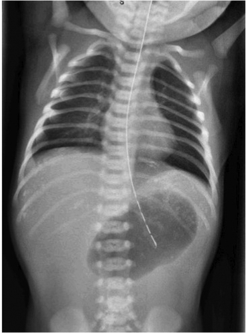
During surgical laparotomy, he was found to have pyloric atresia, antenatal transverse colon perforation, and meconium cyst with meconium peritonitis. His clinical course worsened with secretory diarrhea, intermittent bloody stools, and poor weight gain despite parenteral nutrition and regular albumin infusions.
At 2 months of life, he had persistent feeding difficulties, intermittent fevers without focus of infection, severe normocytic anemia, intermittent leukocytosis, persistent neutrophilia (47 × 109/L), monocytosis (7.2 × 10⁹/L), and lymphopenia (1.3 × 10⁹/L). He had undetectable levels of IgG, IgA, and IgM despite normal albumin levels and a mildly elevated stool alpha-antitrypsin (0.76 g/L, normal 0–0.72 g/L). Upon further evaluation, he had decreased B and CD8 T cell counts: CD19+ 120 × 106 cells/mm (5%), CD4+ 1778 × 106 cells/mm (73%), CD8+ 200 × 106 cells/mm (8%), CD4/CD8 ratio 8.9, CD16_56 268 × 106 cells/mm (11%). B and T lymphocyte phenotyping revealed a high proportion of immature B and T cells (Table 1) with absent switched memory B cells. The lymphocyte stimulation to mitogens was normal (data not shown). A T cell receptor V-beta repertoire showed normal variability without oligoclonal expansion (data not shown).
Table 1:
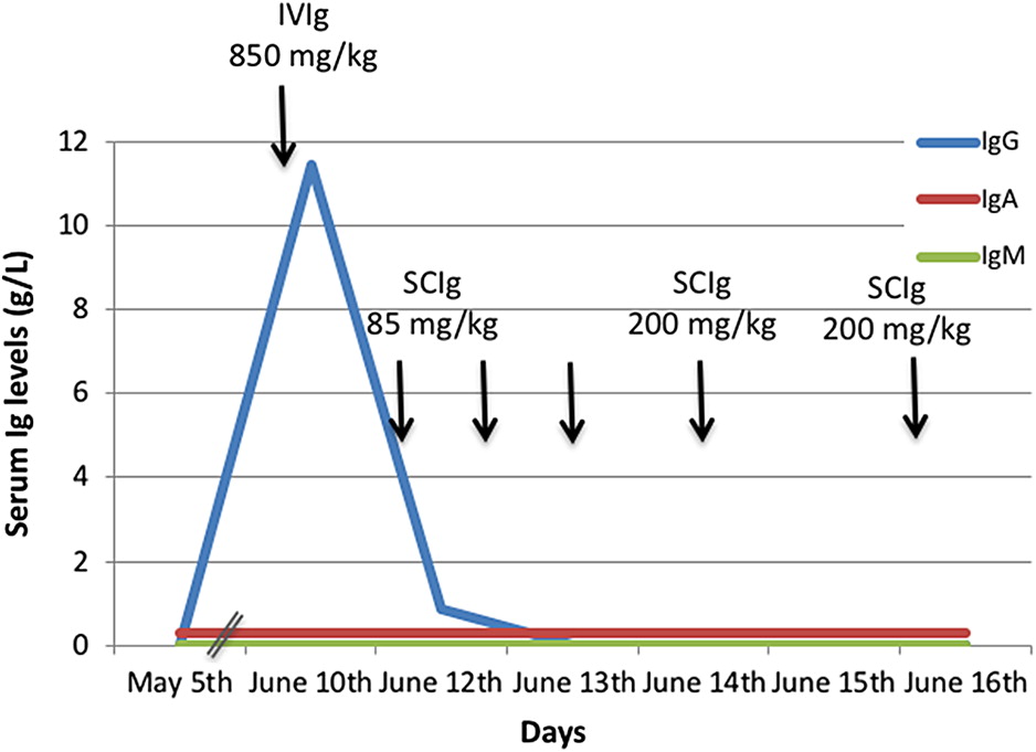
The agammaglobulinemia persisted despite IVIg and subcutaneous immunoglobulin (SCIg) infusion (Figure 2). The IgG level increased to 11.44 g/L (normal range for age 2.22–5.84 g/L) with a loading dose of 850 mg/kg of IVIg but dropped to 0.86 g/L 48 hours later and was undetectable by 72 hours despite daily SCIg doses of 85 mg/kg initiated the day after the IVIg. SCIg doses as high as 200 mg/kg every other day were tried without success. This was attributed to the gastrointestinal losses, although concomitant decreased IgG production remained a possible contributor.
Figure 2:
The infant developed rapidly progressive hepatosplenomegaly and displayed dysautonomic symptoms including hyperthermia, tachycardia, and variable blood pressure. Further investigations including a transesophageal echocardiogram showed subaortic stenosis and moderate mitral regurgitation. He developed clinically significant hypothyroidism at 4 months of age, with a thyroid stimulating hormone (TSH) level as high as 72.39 mIU/L (normal 0.34–5.6 mIU/L). His neonatal screening had previously been reported as normal at 5.37 mIU/L (normal <10 mIU/L) and anti-thyroperoxidase antibodies were absent.
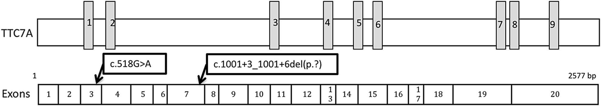
Given his clinical features, gastrointestinal defects and immunodeficiency syndrome (GIDID, MIM 243150) was suspected. Molecular investigations by Next Generation Sequencing revealed compound heterozygous variants in the TTC7A gene (Figure 3). One of those variants, NM_001288951.1:c.1001+3_1001+6del(p.?) causes a frameshift in exon 7 leading to a premature stop codon. This has been reported in the homozygous or compound heterozygous state in French-Canadian patients with multiple intestinal atresia (Chen et al. 2013; Samuels et al. 2013). The second variant, NM_001288951.1:c.518G>A(p. Gly173Asp), had not been previously reported. No other variants including deletion or duplication were detected. Based on his clinical features and the fact that each parent was a heterozygous carrier of one of the two variants identified in the patient, the c.518G>A TTC7A variant is presumed likely causative and pathogenic.
Figure 3:
Given the predictable poor prognosis of this condition, the family elected to pursue with palliative care and declined bowel and hematopoietic stem cell transplantation. In the following weeks, the patient developed worsening respiratory distress and hypoxemia and passed away at 5 months of age.
B cell class switch analysis
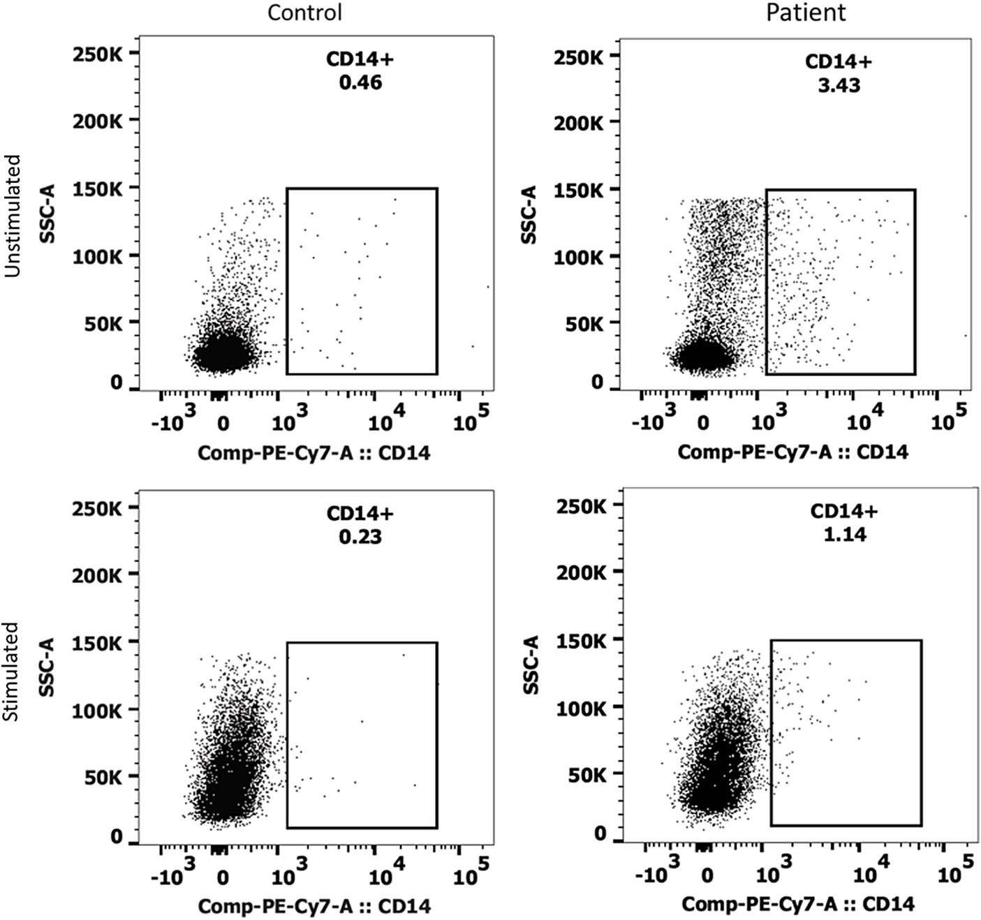
The patient’s PBMCs were cultured for 7 days with or without stimulation with anti-CD40+IL-4+IL-21. At day 7, the microscopic appearance of the patient’s PBMCs appeared very different than control cells, with very few B cell clones and an abundance of dendritic-looking cells (Figure 4). The unstimulated patient’s cells on day 7 had higher CD14+ cells (3.43%) than control (0.46%), which was lower with stimulation at 1.14% and 0.23%, respectively (Figure 5). The day 7 unstimulated patient cells also had a larger CD11b+ cell population (8.62%) that was not present in the control cells (0.27%). Upon stimulation, this CD11b+ population decreased to 2.26% yet remained relatively unchanged in the control. There was a population of double positive CD11b+CD11c+ cells in the unstimulated patient cells, which was also not seen in the control cells (8.85% vs. 0.98%, respectively) (data not shown).
Figure 4:
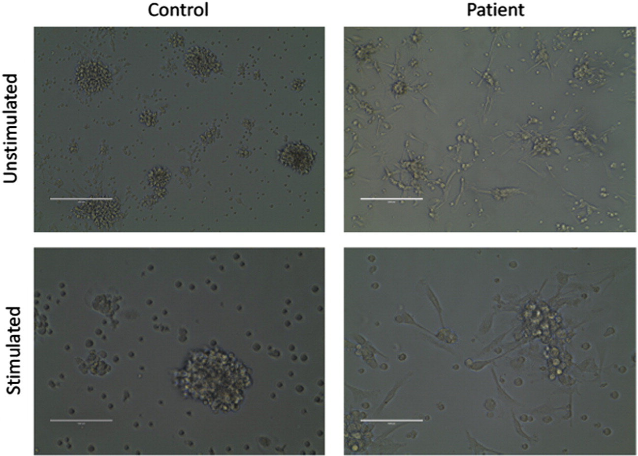
Figure 5:
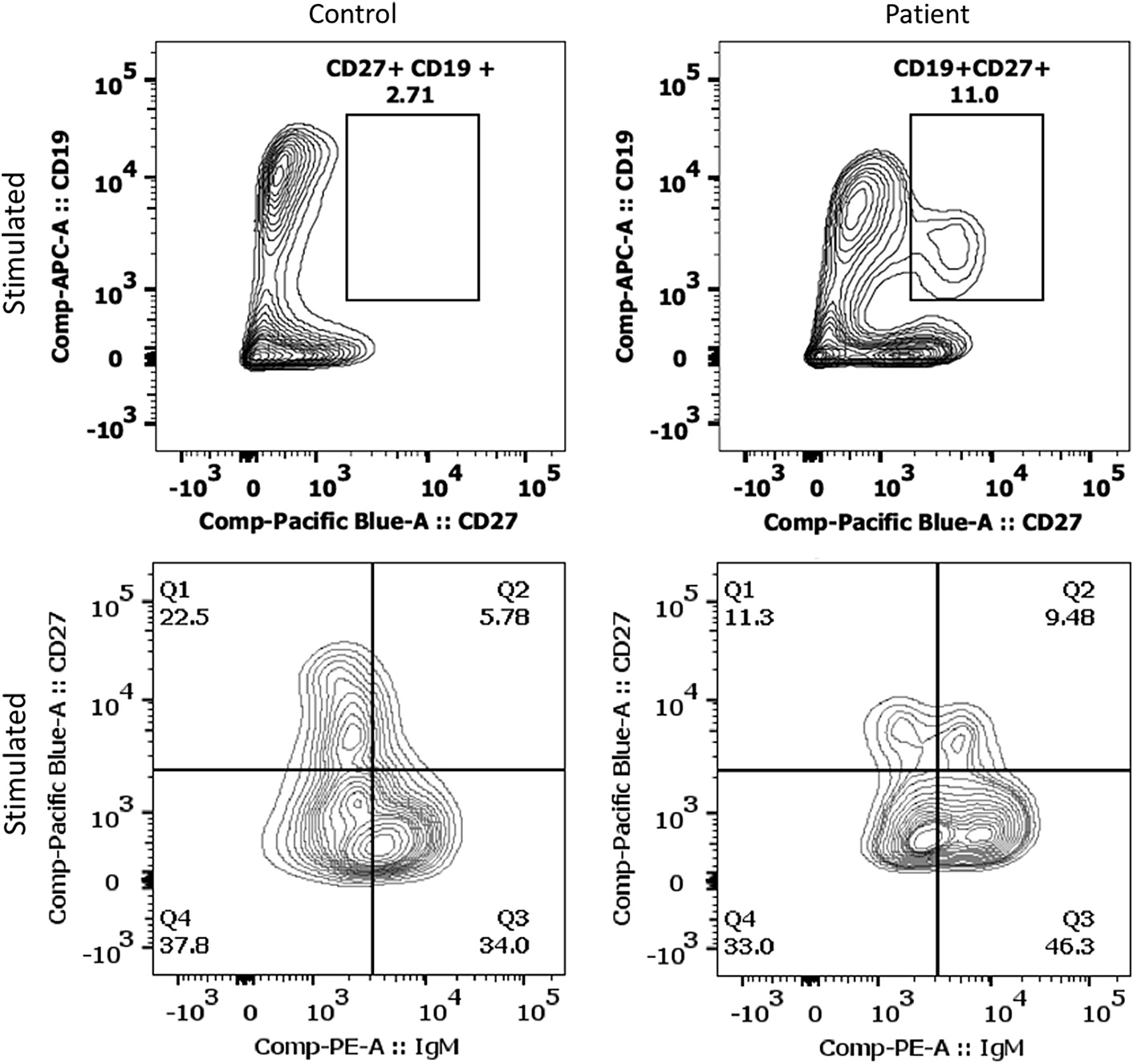
After 7 days of culture, the patient developed a population of CD19+CD27+ B cells that was larger than control (Figure 6). Under stimulation with anti-CD40+IL-4+IL-21, the patient demonstrated 11.0% CD19+CD27+ cells compared to 2.71% in the control. Within the CD19+CD27+ cells, the patient also had a greater CD19+IgM+ population than the control cells (9.48% vs. 5.78%, respectively).
Figure 6:
The supernatant obtained after stimulation with anti-CD40+IL-4+IL-21 at day 7 contained comparable levels of total IgG as the control (755 ng/mL vs. 658 ng/mL, respectively).
Discussion
We report here for the first time a case of GIDID presenting with typical multiple intestinal atresia and combined immunodeficiency but also demonstrating novel features. He presented with acquired hypothyroidism with a TSH level as high as 72.38 mUI/L at 4 months of age, with a previously normal thyroid newborn screen and absence of anti-thyroperoxydase antibodies. He also displayed cardiac anomalies, including subaortic stenosis and moderate mitral regurgitation. To our knowledge, thyroid involvement has not been described in previous patients with GIDID. One paper reported a subject with probable cardiomyopathy (Fernandez et al. 2014) but no clear cardiac malformations. This highlights the multi-systemic involvement of the disease also suggested by previous authors (Fernandez et al. 2014).
Moreover, we present a patient with compound heterozygous mutations with a novel amino acid substitution in TTC7A (Figure 3). The variant NM_001288951.1:c.1001+3_1001+6del(p.?) causes a frameshift in exon 7 leading to a premature stop codon. This has been reported in the homozygous or compound heterozygous state in French-Canadian patients with multiple intestinal atresia (Chen et al. 2013; Samuels et al. 2013). The father is from Bas-du-Fleuve, an area of Canada where a founder effect has been previously described for this TTC7A variant (Samuels et al. 2013). The second variant, NM_001288951.1:c.518G>A(p. Gly173Asp), had not been previously reported. No other variants including deletion or duplication were detected. This rare variant has an extremely low allele frequency of 4.063 × 10⁻⁶ and had been reported only in individuals of Non-Finnish European descent (Lek et al. 2016). The glycine at position 173 is highly conserved across species. It is located within the first tetratricopeptide repeat (TPR) domain of the TTC7A protein. TPR domains are structural motifs important for protein–protein interactions. Based on his clinical features and the fact that each parent was a heterozygous carrier of one of the two variants identified in the patient, the c.518G>A TTC7A variant is presumed likely causative and pathogenic.
Further, our clinical experience and in vitro results suggest that agammaglobulinemia in this case may be due to gastrointestinal loss rather than a defect in antibody production. This patient presented with the classical features of GIDID with significant and secondary immune deficiency. As shown above, he exhibited agammaglobulinemia and displayed in vivo B cell lymphopenia with absent switched memory B cells. Replacement with high doses of SCIg or IVIg was unsuccessful in achieving normal levels of immunoglobulins. This clinically suggests gastrointestinal loss as opposed to a B cell defect.
In vitro studies showed that, under optimal conditions and stimulation, the patient’s B cells were able to produce levels of immunoglobulins comparable to the control when measured in the supernatant obtained after 7 days of culture with anti-CD40+IL-4+IL-21 stimulation. The patient’s PBMCs were compared to an adult control given the inability to obtain an age-matched control. Flow cytometry on day 7 show a larger population of CD19+CD27+ cells in the patient when compared to control (Figure 6). This is in keeping with the hypothesis that the agammaglobulinemia is secondary to the gastrointestinal losses instead of an intrinsic B cell defect in class switching and antibody production. However, the higher number of CD19+CD27+ cells in the patient is probably a reflection of the age difference between the patient and control.
The cellular arm of his immune system was also severely affected with lymphopenia and an abnormal CD4/CD8 ratio of 8.9. This may have been the result of a thymic production defect due to poor cellular polarization, as for the GI epithelium, or preferential gastrointestinal loss of CD8+ T cells.
Conclusion
Our patient presented with a classical and severe clinical picture of GIDID confirmed by compound heterozygous variants in TTC7A. The immunologic phenotype was remarkable for evidence of agammaglobulinemia, decreased B cells and CD8 T cells, defects in lymphocyte maturation, and mildly decreased proliferation to mitogens. Interestingly, under the proper stimulation in vitro, the IgG production was comparable to control. Whether this environment is attainable for the B cells in vivo is unknown. There were other obvious differences in cell populations in our patient compared to control, as seen in the increased number of dendritic cells and macrophages, as well as IgM+ memory B cells. There may not be an inherent defect in class switching and antibody production in B cells, however the physical and cytokine milieu may be defective for cell–cell communication and signaling. Our findings support that the humoral impairment related to TTC7A mutations may be secondary to gastrointestinal loss more than a primary defect. The cellular defects may be more directly related to the TTC7A function. Further studies addressing the function of both the B and T cells are needed in this disease. It is also crucial to assess the role of hematopoietic stem cell transplant, intestinal transplant, and gene therapy in the management of these critically ill patients.
REFERENCES
Avitzur Y., Guo C., Mastropaolo L.A., Bahrami E., Chen H., Zhao Z., Elkadri A., Dhillon S., Murchie R., Fattouh R., Huynh H., Walker J.L., Wales P.W., Cutz E., Kakuta Y., Dudley J., Kammermeier J., Powrie F., Shah N., Walz C., Nathrath M., Kotlarz D., Puchaka J., Krieger J.R., Racek T., Kirchner T., Walters T.D., Brumell J.H., Griffiths A.M., Rezaei N., Rashtian P., Najafi M., Monajemzadeh M., Pelsue S., McGovern D.P., Uhlig H.H., Schadt E., Klein C., Snapper S.B., and Muise A.M. 2014. Mutations in tetratricopeptide repeat domain 7A result in a severe form of very early onset inflammatory bowel disease. Gastroenterology, 146(4):1028–1039.
Bigorgne A.E., Farin H.F., Lemoine R., Mahlaoui N., Lambert N., Gil M., Schulz A., Philippet P., Schlesser P., Abrahamsen T.G., Oymar K., Davies E.G., Ellingsen C.L., Leteurtre E., Moreau-Massart B., Berrebi D., Bole-Feysot C., Nischke P., Brousse N., Fischer A., Clevers H., and de Saint Basile G. 2014. TTC7A mutations disrupt intestinal epithelial apicobasal polarity. J Clin. Invest. 124(1):328–337.
Chen R., Giliani S., Lanzi G., Mias G.I., Lonardi S., Dobbs K., Manis J., Im H., Gallagher J.E., Phanstiel D.H., Euskirchen G., Lacroute P., Bettinger K., Moratto D., Weinacht K., Montin D., Gallo E., Mangili G., Porta F., Notarangelo L.D., Pedretti S., Al-Herz W., Alfahdli W., Comeau A.M., Traister R.S., Pai S.Y., Carella G., Facchetti F., Nadeau K.C., Snyder M., and Notarangelo L.D. 2013. Whole-exome sequencing identifies tetratricopeptide repeat domain 7A (TTC7A) mutations for combined immunodeficiency with intestinal atresias. J. Allergy Clin. Immunol. 132(3):656–664.e17.
Desjardins M., Béland M., Dembele M., Lejtenyi D., Drolet J.P., Lemire M., Tsoukas C., Ben-Shoshan M., Noya F.J.D., Alizadehfar R., McCusker C.T., and Mazer B.D. 2018. Modulation of the interleukin-21 pathway with interleukin-4 distinguishes common variable immunodeficiency patients with more non-infectious clinical complications. J. Clin. Immunol. 38(1):45–55.
Fernandez I., Patey N., Marchand V., Birlea M., Maranda B., Haddad E., Decaluwe H., and Le Deist F. 2014. Multiple intestinal atresia with combined immune deficiency related to TTC7A defect is a multiorgan pathology: Study of a French-Canadian-based cohort. Medicine, 93(29):e327.
Leclerc-Mercier S., Lemoine R., Bigorgne A.E., Sepulveda F., Leveau C., Fischer A., Mahlaoui N., Hadj-Rabia S., and de Saint Basile G. 2016. Ichthyosis as the dermatological phenotype associated with TTC7A mutations. Br. J. Dermatol. 175(5):1061–1064.
Lek M., Karczewski K.J., Minikel E.V., Samocha K.E., Banks E., Fennell T., O’Donnell-Luria A.H., Ware J.S., Hill A.J., Cummings B.B., Tukiainen T., Birnbaum D.P., Kosmicki J.A., Duncan L.E., Estrada K., Zhao F., Zou J., Pierce-Hoffman E., Berghout J., Cooper D.N., Deflaux N., DePristo M., Do R., Flannick J., Fromer M., Gauthier L., Goldstein J., Gupta N., Howrigan D., Kiezun A., Kurki M.I., Moonshine A.L., Natarajan P., Orozco L., Peloso G.M., Poplin R., Rivas M.A., Ruano-Rubio V., Rose S.A., Ruderfer D.M., Shakir K., Stenson P.D., Stevens C., Thomas B.P., Tiao G., Tusie-Luna M.T., Weisburd B., Won H.H., Yu D., Altshuler D.M., Ardissino D., Boehnke M., Danesh J., Donnelly S., Elosua R., Florez J.C., Gabriel S.B., Getz G., Glatt S.J., Hultman C.M., Kathiresan S., Laakso M., McCarroll S., McCarthy M.I., McGovern D., McPherson R., Neale B.M., Palotie A., Purcell S.M., Saleheen D., Scharf J.M., Sklar P., Sullivan P.F., Tuomilehto J., Tsuang M.T., Watkins H.C., Wilson J.G., Daly M.J., MacArthur D.G., and and Exome Aggregation Consortium 2016. Analysis of protein-coding genetic variation in 60,706 humans. Nature, 536(7616):285–291.
Lemoine R., Bigorgne A., Farin H., and Saint Basile Gd. 2014a. TTC7A, a critical effector for the intestinal and immune system homeostasis. Med. Sci. 30(6–7):616–618.
Lemoine R., Pachlopnik-schmid J., Pachlopnik-Schmid J., Farin H.F., Bigorgne A., Debré M., Sepulveda F., Héritier S., Lemale J., Talbotec C., Rieux-Laucat F., Ruemmele F., Morali A., Cathebras P., Nitschke P., Bole-Feysot C., Blanche S., Brousse N., Picard C., Clevers H., Fischer A., and de Saint Basile G. 2014b. Immune deficiency-related enteropathy-lymphocytopenia-alopecia syndrome results from tetratricopeptide repeat domain 7A deficiency. J Allergy Clin. Immunol. 134(6):1354–1364.e6.
Ngan, B., Merico, D., Marcus, N., Kim, V.H.D., Upton, J., Bates, A., Herbrick, J., Nalpathamkalam, T., Thiruvahindrapuram, B., Cox, P., and Roifman, C.M. 2014. Mutations in tetratricopeptide repeat domain 7A (TTC7A) are associated with combined immunodeficiency with dendriform lung ossification but no intestinal atresia. LymphoSign J. 1(1):10–26. [Online]. Available from http://lymphosign.com/doi/abs/10.14785/lpsn-2014-0002.
Samuels M.E., Majewski J., Alirezaie N., Fernandez I., Casals F., Patey N., Decaluwe H., Gosselin I., Haddad E., Hodgkinson A., Idaghdour Y., Marchand V., Michaud J.L., Rodrigue M.A., Desjardins S., Dubois S., Le Deist F., Awadalla P., Raymond V., and Maranda B. 2013. Exome sequencing identifies mutations in the gene TTC7A in French-Canadian cases with hereditary multiple intestinal atresia. J. Med. Genet. 50(5):324–329.
Information & Authors
Information
Published In
LymphoSign Journal
Volume 5 • Number 3 • September 2018
Pages: 91 - 99
History
Received: 1 July 2018
Accepted: 23 July 2018
Accepted manuscript online: 16 August 2018
Copyright
© 2018.
Authors
Metrics & Citations
Metrics
Other Metrics
Citations
Cite As
AlexandraLanglois, BaharTorabi, MariemeDembele, MarylinDesjardins, RezaAlizadehfar, MosheBen-Shohan, IsabelleDe Bie, AnaSantanna, ChristineMcCusker, and BruceMazer. 2018. Gastrointestinal defects and immunodeficiency syndrome with normal in vitro IgG production. LymphoSign Journal.
5(3): 91-99. https://doi.org/10.14785/lymphosign-2018-0008
Export Citations
If you have the appropriate software installed, you can download article citation data to the citation manager of your choice. Simply select your manager software from the list below and click Download.
There are no citations for this item
View Options
View options
Get Access
Login options
Check if you access through your login credentials or your institution to get full access on this article.
